Rohollah Nasiri, Myra Kurosu Jalil, Veronica Ibanez Gaspar, Andrea Sofia Flores Perez, Hieu Thi Minh Nguyen, Syamantak Khan, Sindy K. Y. Tang, Yunzhi Peter Yang and Guillem Pratx
{"title":"一个肺肿瘤芯片模型概括了缺氧对放疗反应和FDG-PET成像的影响","authors":"Rohollah Nasiri, Myra Kurosu Jalil, Veronica Ibanez Gaspar, Andrea Sofia Flores Perez, Hieu Thi Minh Nguyen, Syamantak Khan, Sindy K. Y. Tang, Yunzhi Peter Yang and Guillem Pratx","doi":"10.1039/D5LC00373C","DOIUrl":null,"url":null,"abstract":"<p >Most solid tumors contain regions of hypoxia that pose a significant challenge to the efficacy of radiation therapy. This study introduces a novel 3D lung tumor-on-a-chip (ToC) model designed to replicate the hypoxic tumor microenvironment <em>in vitro</em> while also providing a platform for clinically relevant interventions such as radiotherapy and positron emission tomography (PET) imaging. To simulate the heterogeneous oxygen distribution found in tumors, the ToC model incorporates an oxygen gradient achieved through a straightforward chemical oxygen scavenging system. A unique innovation of this device is the integration of a thin scintillator plate, which enables high-resolution radioluminescence microscopy imaging of tumor metabolism under hypoxia and normoxia conditions using clinically approved PET tracers such as fluorodeoxyglucose (FDG). The response of this hypoxic model to radiation therapy (10 Gy X-ray) demonstrated ∼4-fold higher radioresistance compared to the normoxic ToC model, as assessed by colony formation potential. Additionally, DNA damage observed in the normoxic ToC model was ∼5-fold higher than that in the hypoxic model. Furthermore, the metabolic consumption of glucose was found to mirror the localization of hypoxia, validating the use of this biomarker for planning radiation therapy. The integration of high-resolution radionuclide imaging within ToC models enables on-chip PET imaging and facilitates oncology research and discovery, offering innovative capabilities for the preclinical testing of novel cancer therapies in a clinically relevant environment.</p>","PeriodicalId":85,"journal":{"name":"Lab on a Chip","volume":" 18","pages":" 4677-4691"},"PeriodicalIF":5.4000,"publicationDate":"2025-07-30","publicationTypes":"Journal Article","fieldsOfStudy":null,"isOpenAccess":false,"openAccessPdf":"","citationCount":"0","resultStr":"{\"title\":\"A lung tumor-on-a-chip model recapitulates the effect of hypoxia on radiotherapy response and FDG-PET imaging\",\"authors\":\"Rohollah Nasiri, Myra Kurosu Jalil, Veronica Ibanez Gaspar, Andrea Sofia Flores Perez, Hieu Thi Minh Nguyen, Syamantak Khan, Sindy K. Y. Tang, Yunzhi Peter Yang and Guillem Pratx\",\"doi\":\"10.1039/D5LC00373C\",\"DOIUrl\":null,\"url\":null,\"abstract\":\"<p >Most solid tumors contain regions of hypoxia that pose a significant challenge to the efficacy of radiation therapy. This study introduces a novel 3D lung tumor-on-a-chip (ToC) model designed to replicate the hypoxic tumor microenvironment <em>in vitro</em> while also providing a platform for clinically relevant interventions such as radiotherapy and positron emission tomography (PET) imaging. To simulate the heterogeneous oxygen distribution found in tumors, the ToC model incorporates an oxygen gradient achieved through a straightforward chemical oxygen scavenging system. A unique innovation of this device is the integration of a thin scintillator plate, which enables high-resolution radioluminescence microscopy imaging of tumor metabolism under hypoxia and normoxia conditions using clinically approved PET tracers such as fluorodeoxyglucose (FDG). The response of this hypoxic model to radiation therapy (10 Gy X-ray) demonstrated ∼4-fold higher radioresistance compared to the normoxic ToC model, as assessed by colony formation potential. Additionally, DNA damage observed in the normoxic ToC model was ∼5-fold higher than that in the hypoxic model. Furthermore, the metabolic consumption of glucose was found to mirror the localization of hypoxia, validating the use of this biomarker for planning radiation therapy. The integration of high-resolution radionuclide imaging within ToC models enables on-chip PET imaging and facilitates oncology research and discovery, offering innovative capabilities for the preclinical testing of novel cancer therapies in a clinically relevant environment.</p>\",\"PeriodicalId\":85,\"journal\":{\"name\":\"Lab on a Chip\",\"volume\":\" 18\",\"pages\":\" 4677-4691\"},\"PeriodicalIF\":5.4000,\"publicationDate\":\"2025-07-30\",\"publicationTypes\":\"Journal Article\",\"fieldsOfStudy\":null,\"isOpenAccess\":false,\"openAccessPdf\":\"\",\"citationCount\":\"0\",\"resultStr\":null,\"platform\":\"Semanticscholar\",\"paperid\":null,\"PeriodicalName\":\"Lab on a Chip\",\"FirstCategoryId\":\"5\",\"ListUrlMain\":\"https://pubs.rsc.org/en/content/articlelanding/2025/lc/d5lc00373c\",\"RegionNum\":2,\"RegionCategory\":\"工程技术\",\"ArticlePicture\":[],\"TitleCN\":null,\"AbstractTextCN\":null,\"PMCID\":null,\"EPubDate\":\"\",\"PubModel\":\"\",\"JCR\":\"Q1\",\"JCRName\":\"BIOCHEMICAL RESEARCH METHODS\",\"Score\":null,\"Total\":0}","platform":"Semanticscholar","paperid":null,"PeriodicalName":"Lab on a Chip","FirstCategoryId":"5","ListUrlMain":"https://pubs.rsc.org/en/content/articlelanding/2025/lc/d5lc00373c","RegionNum":2,"RegionCategory":"工程技术","ArticlePicture":[],"TitleCN":null,"AbstractTextCN":null,"PMCID":null,"EPubDate":"","PubModel":"","JCR":"Q1","JCRName":"BIOCHEMICAL RESEARCH METHODS","Score":null,"Total":0}
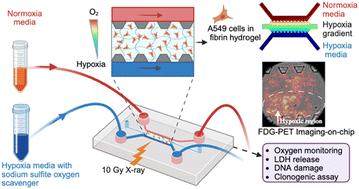
A lung tumor-on-a-chip model recapitulates the effect of hypoxia on radiotherapy response and FDG-PET imaging
Most solid tumors contain regions of hypoxia that pose a significant challenge to the efficacy of radiation therapy. This study introduces a novel 3D lung tumor-on-a-chip (ToC) model designed to replicate the hypoxic tumor microenvironment in vitro while also providing a platform for clinically relevant interventions such as radiotherapy and positron emission tomography (PET) imaging. To simulate the heterogeneous oxygen distribution found in tumors, the ToC model incorporates an oxygen gradient achieved through a straightforward chemical oxygen scavenging system. A unique innovation of this device is the integration of a thin scintillator plate, which enables high-resolution radioluminescence microscopy imaging of tumor metabolism under hypoxia and normoxia conditions using clinically approved PET tracers such as fluorodeoxyglucose (FDG). The response of this hypoxic model to radiation therapy (10 Gy X-ray) demonstrated ∼4-fold higher radioresistance compared to the normoxic ToC model, as assessed by colony formation potential. Additionally, DNA damage observed in the normoxic ToC model was ∼5-fold higher than that in the hypoxic model. Furthermore, the metabolic consumption of glucose was found to mirror the localization of hypoxia, validating the use of this biomarker for planning radiation therapy. The integration of high-resolution radionuclide imaging within ToC models enables on-chip PET imaging and facilitates oncology research and discovery, offering innovative capabilities for the preclinical testing of novel cancer therapies in a clinically relevant environment.
期刊介绍:
Lab on a Chip is the premiere journal that publishes cutting-edge research in the field of miniaturization. By their very nature, microfluidic/nanofluidic/miniaturized systems are at the intersection of disciplines, spanning fundamental research to high-end application, which is reflected by the broad readership of the journal. Lab on a Chip publishes two types of papers on original research: full-length research papers and communications. Papers should demonstrate innovations, which can come from technical advancements or applications addressing pressing needs in globally important areas. The journal also publishes Comments, Reviews, and Perspectives.
 求助内容:
求助内容: 应助结果提醒方式:


