Anastasiia Asmolova, Anastasiia Sukmanova, Milana Makarova, Pavel Novikov, Vadim Nikulin, Maria Nazarova
{"title":"刺激间隔与多肌nTMS运动映射关系的研究。","authors":"Anastasiia Asmolova, Anastasiia Sukmanova, Milana Makarova, Pavel Novikov, Vadim Nikulin, Maria Nazarova","doi":"10.1007/s10548-025-01128-9","DOIUrl":null,"url":null,"abstract":"<p><p>Although the interstimulus interval (ISI) is one of the crucial parameters in the transcranial magnetic stimulation (TMS), the ISI effect on the results of the TMS motor mapping is usually overlooked. This study explored the influence of ISI, ranging from 1.5 to 41 s, on multi-muscle navigated TMS (nTMS) motor mapping results. Twenty-six healthy male volunteers underwent four nTMS motor mapping sessions on two separate days. We mapped the muscles' cortical representations (MCRs) of the five upper limb muscles: abductor pollicis brevis (APB), abductor digiti minimi (ADM), first dorsal interosseous (FDI), extensor digitorum communis (EDC), and biceps brachii (BB). We estimated the relationship between ISIs and trial-to-trial motor evoked potentials (MEPs) amplitudes and MCR areas. In addition, we accounted for the association between the ISI and TMS mapping procedure parameters such as the distance between the successive stimulation points, the number of stimuli in a TMS session, and the stimulus counting number. A weak positive association was observed between: (1) trial-to-trial ISI and MEP amplitude and (2) median ISI and MCR areas. We recommend reporting ISI values in TMS motor mapping studies and monitoring the impact of ISI on MEP amplitudes.</p>","PeriodicalId":55329,"journal":{"name":"Brain Topography","volume":"38 5","pages":"55"},"PeriodicalIF":2.9000,"publicationDate":"2025-07-30","publicationTypes":"Journal Article","fieldsOfStudy":null,"isOpenAccess":false,"openAccessPdf":"https://www.ncbi.nlm.nih.gov/pmc/articles/PMC12310799/pdf/","citationCount":"0","resultStr":"{\"title\":\"On the Relation Between the Interstimulus Intervals and Multi-Muscle nTMS Motor Mapping.\",\"authors\":\"Anastasiia Asmolova, Anastasiia Sukmanova, Milana Makarova, Pavel Novikov, Vadim Nikulin, Maria Nazarova\",\"doi\":\"10.1007/s10548-025-01128-9\",\"DOIUrl\":null,\"url\":null,\"abstract\":\"<p><p>Although the interstimulus interval (ISI) is one of the crucial parameters in the transcranial magnetic stimulation (TMS), the ISI effect on the results of the TMS motor mapping is usually overlooked. This study explored the influence of ISI, ranging from 1.5 to 41 s, on multi-muscle navigated TMS (nTMS) motor mapping results. Twenty-six healthy male volunteers underwent four nTMS motor mapping sessions on two separate days. We mapped the muscles' cortical representations (MCRs) of the five upper limb muscles: abductor pollicis brevis (APB), abductor digiti minimi (ADM), first dorsal interosseous (FDI), extensor digitorum communis (EDC), and biceps brachii (BB). We estimated the relationship between ISIs and trial-to-trial motor evoked potentials (MEPs) amplitudes and MCR areas. In addition, we accounted for the association between the ISI and TMS mapping procedure parameters such as the distance between the successive stimulation points, the number of stimuli in a TMS session, and the stimulus counting number. A weak positive association was observed between: (1) trial-to-trial ISI and MEP amplitude and (2) median ISI and MCR areas. We recommend reporting ISI values in TMS motor mapping studies and monitoring the impact of ISI on MEP amplitudes.</p>\",\"PeriodicalId\":55329,\"journal\":{\"name\":\"Brain Topography\",\"volume\":\"38 5\",\"pages\":\"55\"},\"PeriodicalIF\":2.9000,\"publicationDate\":\"2025-07-30\",\"publicationTypes\":\"Journal Article\",\"fieldsOfStudy\":null,\"isOpenAccess\":false,\"openAccessPdf\":\"https://www.ncbi.nlm.nih.gov/pmc/articles/PMC12310799/pdf/\",\"citationCount\":\"0\",\"resultStr\":null,\"platform\":\"Semanticscholar\",\"paperid\":null,\"PeriodicalName\":\"Brain Topography\",\"FirstCategoryId\":\"3\",\"ListUrlMain\":\"https://doi.org/10.1007/s10548-025-01128-9\",\"RegionNum\":3,\"RegionCategory\":\"医学\",\"ArticlePicture\":[],\"TitleCN\":null,\"AbstractTextCN\":null,\"PMCID\":null,\"EPubDate\":\"\",\"PubModel\":\"\",\"JCR\":\"Q3\",\"JCRName\":\"CLINICAL NEUROLOGY\",\"Score\":null,\"Total\":0}","platform":"Semanticscholar","paperid":null,"PeriodicalName":"Brain Topography","FirstCategoryId":"3","ListUrlMain":"https://doi.org/10.1007/s10548-025-01128-9","RegionNum":3,"RegionCategory":"医学","ArticlePicture":[],"TitleCN":null,"AbstractTextCN":null,"PMCID":null,"EPubDate":"","PubModel":"","JCR":"Q3","JCRName":"CLINICAL NEUROLOGY","Score":null,"Total":0}
On the Relation Between the Interstimulus Intervals and Multi-Muscle nTMS Motor Mapping.
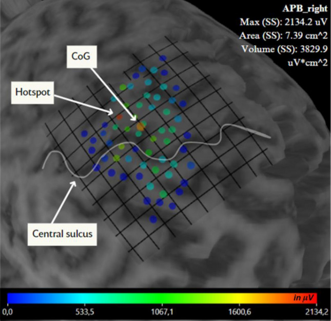
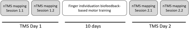
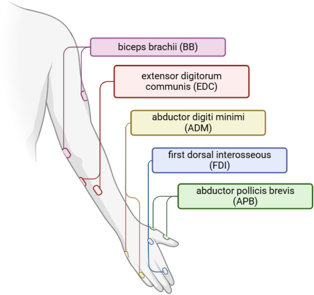
Although the interstimulus interval (ISI) is one of the crucial parameters in the transcranial magnetic stimulation (TMS), the ISI effect on the results of the TMS motor mapping is usually overlooked. This study explored the influence of ISI, ranging from 1.5 to 41 s, on multi-muscle navigated TMS (nTMS) motor mapping results. Twenty-six healthy male volunteers underwent four nTMS motor mapping sessions on two separate days. We mapped the muscles' cortical representations (MCRs) of the five upper limb muscles: abductor pollicis brevis (APB), abductor digiti minimi (ADM), first dorsal interosseous (FDI), extensor digitorum communis (EDC), and biceps brachii (BB). We estimated the relationship between ISIs and trial-to-trial motor evoked potentials (MEPs) amplitudes and MCR areas. In addition, we accounted for the association between the ISI and TMS mapping procedure parameters such as the distance between the successive stimulation points, the number of stimuli in a TMS session, and the stimulus counting number. A weak positive association was observed between: (1) trial-to-trial ISI and MEP amplitude and (2) median ISI and MCR areas. We recommend reporting ISI values in TMS motor mapping studies and monitoring the impact of ISI on MEP amplitudes.
期刊介绍:
Brain Topography publishes clinical and basic research on cognitive neuroscience and functional neurophysiology using the full range of imaging techniques including EEG, MEG, fMRI, TMS, diffusion imaging, spectroscopy, intracranial recordings, lesion studies, and related methods. Submissions combining multiple techniques are particularly encouraged, as well as reports of new and innovative methodologies.
 求助内容:
求助内容: 应助结果提醒方式:


