Raoul Orvieto, Omri Nayshool, Louisa Cohen, Yuval Yung, Adva Aizer, Efrat Glick Saar, Dan Dominissini
{"title":"衰老对人类卵母细胞基因表达的影响:对年轻和老年患者的比较分析。","authors":"Raoul Orvieto, Omri Nayshool, Louisa Cohen, Yuval Yung, Adva Aizer, Efrat Glick Saar, Dan Dominissini","doi":"10.1186/s12958-025-01449-1","DOIUrl":null,"url":null,"abstract":"<p><strong>Background: </strong>Aging affects gene expression in pathways essential for energy metabolism, DNA repair, cell cycle regulation, and antioxidant defenses, directly affecting oocyte quality and viability. Single-cell RNA deep sequencing studies of aged versus young human MII oocytes revealed many differentially expressed genes. In addition, single human oocyte transcriptome analysis at both germinal vesicle (GV) and MII stages revealed distinct stage-dependent pathways impacted by aging, with a decrease in mitochondrial-related transcripts from GV to MII oocytes, and a much greater reduction in MII oocytes with advanced age.</p><p><strong>Objective: </strong>Our aim was to investigate the age-related differences in gene expression of germinal vesicle (GV) oocytes between young and advanced age patients.</p><p><strong>Patients and methods: </strong>Immature GV oocytes were donated by 6 patients, divided into two age groups: The \"Young\" group (ages 16-29) had three participants (mean age: 23.3 ± 6.6 years), and the \"Elderly\" group (ages 38-40) included three participants (mean age: 39 ± 1 year). After retrieval, oocytes were denuded and donated GV oocytes were cryopreserved at -196<sup>0</sup>C until analysis. For library preparation, we used the NEBNext<sup>®</sup> Single Cell/Low Input RNA Library Prep Kit for Illumina, Sect. 1 (cat no. E6420S, New England Biolabs (NEB), USA), strictly adhering to the manufacturer's instructions. Gene expression quantification was performed using feature Counts from the Subread package (v1.5.3), and comprehensive quality control reports were generated using MultiQC (v1.25.1). To further corroborate the differential expression of hub genes associated with oocyte aging identified in our preliminary analysis, quantitative real-time PCR (qPCR) was performed for four selected hub genes (MYL4, POMZP3, and LINC002087).</p><p><strong>Results: </strong>Of top 10 significantly differently expressed genes 7 (LINC02087, POMZP3, LINC02749, MYL4, AGPAT2, GCA, and LIMK1) were downregulated and 3 (CLEC3A, ARPP21, and CITED2) showed significant upregulation in young versus old oocytes. These genes underscore the impact of aging on critical oocyte pathways, including chromosomal stability, epigenetic regulation, mitochondrial function, immune response, structural integrity, and calcium signaling. Moreover, among these genes, LINC02087 was the most downregulated (log2FC = -7.66), while CITED2 showed the strongest upregulation (log2FC = 3.43) in young versus old oocytes. Following the RNA extraction of pooled GV oocytes of 8 elderly and 9 young donors' GV oocytes. We observed significant differences in gene expression levels between the two age groups, in line with the single-cell RNASeq.</p><p><strong>Conclusion: </strong>Understanding the effects of aging on the oocyte transcriptome could identify biomarkers that characterize good MII oocyte quality. The different genes expressions in aged oocytes highlight their potential contributions to oocyte quality and development. Moreover, by elucidating age-related changes across diverse cellular functions, this preliminary study opens avenues for therapeutic interventions aimed at extending reproductive longevity and optimizing outcomes in assisted reproductive technologies.</p>","PeriodicalId":21011,"journal":{"name":"Reproductive Biology and Endocrinology","volume":"23 1","pages":"111"},"PeriodicalIF":4.7000,"publicationDate":"2025-07-29","publicationTypes":"Journal Article","fieldsOfStudy":null,"isOpenAccess":false,"openAccessPdf":"https://www.ncbi.nlm.nih.gov/pmc/articles/PMC12305945/pdf/","citationCount":"0","resultStr":"{\"title\":\"Impact of aging on gene expression in human oocytes: a comparative analysis of young and older patients.\",\"authors\":\"Raoul Orvieto, Omri Nayshool, Louisa Cohen, Yuval Yung, Adva Aizer, Efrat Glick Saar, Dan Dominissini\",\"doi\":\"10.1186/s12958-025-01449-1\",\"DOIUrl\":null,\"url\":null,\"abstract\":\"<p><strong>Background: </strong>Aging affects gene expression in pathways essential for energy metabolism, DNA repair, cell cycle regulation, and antioxidant defenses, directly affecting oocyte quality and viability. Single-cell RNA deep sequencing studies of aged versus young human MII oocytes revealed many differentially expressed genes. In addition, single human oocyte transcriptome analysis at both germinal vesicle (GV) and MII stages revealed distinct stage-dependent pathways impacted by aging, with a decrease in mitochondrial-related transcripts from GV to MII oocytes, and a much greater reduction in MII oocytes with advanced age.</p><p><strong>Objective: </strong>Our aim was to investigate the age-related differences in gene expression of germinal vesicle (GV) oocytes between young and advanced age patients.</p><p><strong>Patients and methods: </strong>Immature GV oocytes were donated by 6 patients, divided into two age groups: The \\\"Young\\\" group (ages 16-29) had three participants (mean age: 23.3 ± 6.6 years), and the \\\"Elderly\\\" group (ages 38-40) included three participants (mean age: 39 ± 1 year). After retrieval, oocytes were denuded and donated GV oocytes were cryopreserved at -196<sup>0</sup>C until analysis. For library preparation, we used the NEBNext<sup>®</sup> Single Cell/Low Input RNA Library Prep Kit for Illumina, Sect. 1 (cat no. E6420S, New England Biolabs (NEB), USA), strictly adhering to the manufacturer's instructions. Gene expression quantification was performed using feature Counts from the Subread package (v1.5.3), and comprehensive quality control reports were generated using MultiQC (v1.25.1). To further corroborate the differential expression of hub genes associated with oocyte aging identified in our preliminary analysis, quantitative real-time PCR (qPCR) was performed for four selected hub genes (MYL4, POMZP3, and LINC002087).</p><p><strong>Results: </strong>Of top 10 significantly differently expressed genes 7 (LINC02087, POMZP3, LINC02749, MYL4, AGPAT2, GCA, and LIMK1) were downregulated and 3 (CLEC3A, ARPP21, and CITED2) showed significant upregulation in young versus old oocytes. These genes underscore the impact of aging on critical oocyte pathways, including chromosomal stability, epigenetic regulation, mitochondrial function, immune response, structural integrity, and calcium signaling. Moreover, among these genes, LINC02087 was the most downregulated (log2FC = -7.66), while CITED2 showed the strongest upregulation (log2FC = 3.43) in young versus old oocytes. Following the RNA extraction of pooled GV oocytes of 8 elderly and 9 young donors' GV oocytes. We observed significant differences in gene expression levels between the two age groups, in line with the single-cell RNASeq.</p><p><strong>Conclusion: </strong>Understanding the effects of aging on the oocyte transcriptome could identify biomarkers that characterize good MII oocyte quality. The different genes expressions in aged oocytes highlight their potential contributions to oocyte quality and development. Moreover, by elucidating age-related changes across diverse cellular functions, this preliminary study opens avenues for therapeutic interventions aimed at extending reproductive longevity and optimizing outcomes in assisted reproductive technologies.</p>\",\"PeriodicalId\":21011,\"journal\":{\"name\":\"Reproductive Biology and Endocrinology\",\"volume\":\"23 1\",\"pages\":\"111\"},\"PeriodicalIF\":4.7000,\"publicationDate\":\"2025-07-29\",\"publicationTypes\":\"Journal Article\",\"fieldsOfStudy\":null,\"isOpenAccess\":false,\"openAccessPdf\":\"https://www.ncbi.nlm.nih.gov/pmc/articles/PMC12305945/pdf/\",\"citationCount\":\"0\",\"resultStr\":null,\"platform\":\"Semanticscholar\",\"paperid\":null,\"PeriodicalName\":\"Reproductive Biology and Endocrinology\",\"FirstCategoryId\":\"3\",\"ListUrlMain\":\"https://doi.org/10.1186/s12958-025-01449-1\",\"RegionNum\":2,\"RegionCategory\":\"医学\",\"ArticlePicture\":[],\"TitleCN\":null,\"AbstractTextCN\":null,\"PMCID\":null,\"EPubDate\":\"\",\"PubModel\":\"\",\"JCR\":\"Q1\",\"JCRName\":\"ENDOCRINOLOGY & METABOLISM\",\"Score\":null,\"Total\":0}","platform":"Semanticscholar","paperid":null,"PeriodicalName":"Reproductive Biology and Endocrinology","FirstCategoryId":"3","ListUrlMain":"https://doi.org/10.1186/s12958-025-01449-1","RegionNum":2,"RegionCategory":"医学","ArticlePicture":[],"TitleCN":null,"AbstractTextCN":null,"PMCID":null,"EPubDate":"","PubModel":"","JCR":"Q1","JCRName":"ENDOCRINOLOGY & METABOLISM","Score":null,"Total":0}
Impact of aging on gene expression in human oocytes: a comparative analysis of young and older patients.
Background: Aging affects gene expression in pathways essential for energy metabolism, DNA repair, cell cycle regulation, and antioxidant defenses, directly affecting oocyte quality and viability. Single-cell RNA deep sequencing studies of aged versus young human MII oocytes revealed many differentially expressed genes. In addition, single human oocyte transcriptome analysis at both germinal vesicle (GV) and MII stages revealed distinct stage-dependent pathways impacted by aging, with a decrease in mitochondrial-related transcripts from GV to MII oocytes, and a much greater reduction in MII oocytes with advanced age.
Objective: Our aim was to investigate the age-related differences in gene expression of germinal vesicle (GV) oocytes between young and advanced age patients.
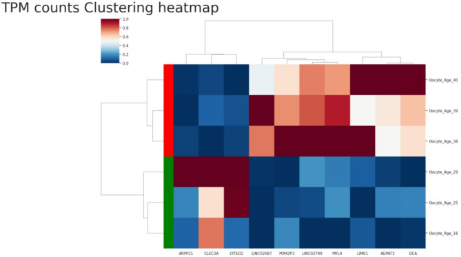
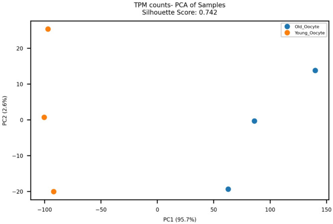
Patients and methods: Immature GV oocytes were donated by 6 patients, divided into two age groups: The "Young" group (ages 16-29) had three participants (mean age: 23.3 ± 6.6 years), and the "Elderly" group (ages 38-40) included three participants (mean age: 39 ± 1 year). After retrieval, oocytes were denuded and donated GV oocytes were cryopreserved at -1960C until analysis. For library preparation, we used the NEBNext® Single Cell/Low Input RNA Library Prep Kit for Illumina, Sect. 1 (cat no. E6420S, New England Biolabs (NEB), USA), strictly adhering to the manufacturer's instructions. Gene expression quantification was performed using feature Counts from the Subread package (v1.5.3), and comprehensive quality control reports were generated using MultiQC (v1.25.1). To further corroborate the differential expression of hub genes associated with oocyte aging identified in our preliminary analysis, quantitative real-time PCR (qPCR) was performed for four selected hub genes (MYL4, POMZP3, and LINC002087).
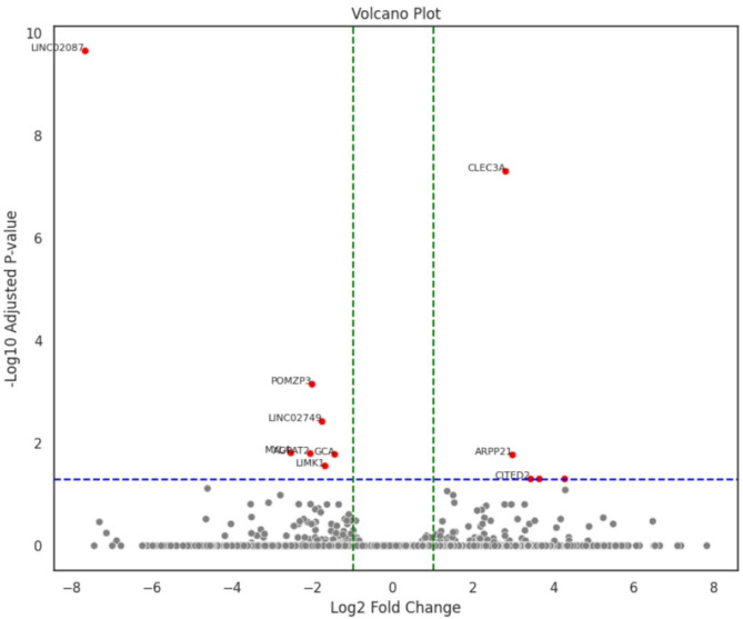
Results: Of top 10 significantly differently expressed genes 7 (LINC02087, POMZP3, LINC02749, MYL4, AGPAT2, GCA, and LIMK1) were downregulated and 3 (CLEC3A, ARPP21, and CITED2) showed significant upregulation in young versus old oocytes. These genes underscore the impact of aging on critical oocyte pathways, including chromosomal stability, epigenetic regulation, mitochondrial function, immune response, structural integrity, and calcium signaling. Moreover, among these genes, LINC02087 was the most downregulated (log2FC = -7.66), while CITED2 showed the strongest upregulation (log2FC = 3.43) in young versus old oocytes. Following the RNA extraction of pooled GV oocytes of 8 elderly and 9 young donors' GV oocytes. We observed significant differences in gene expression levels between the two age groups, in line with the single-cell RNASeq.
Conclusion: Understanding the effects of aging on the oocyte transcriptome could identify biomarkers that characterize good MII oocyte quality. The different genes expressions in aged oocytes highlight their potential contributions to oocyte quality and development. Moreover, by elucidating age-related changes across diverse cellular functions, this preliminary study opens avenues for therapeutic interventions aimed at extending reproductive longevity and optimizing outcomes in assisted reproductive technologies.
期刊介绍:
Reproductive Biology and Endocrinology publishes and disseminates high-quality results from excellent research in the reproductive sciences.
The journal publishes on topics covering gametogenesis, fertilization, early embryonic development, embryo-uterus interaction, reproductive development, pregnancy, uterine biology, endocrinology of reproduction, control of reproduction, reproductive immunology, neuroendocrinology, and veterinary and human reproductive medicine, including all vertebrate species.
 求助内容:
求助内容: 应助结果提醒方式:


