{"title":"免疫复合物介导的膜性增殖性肾小球肾炎III型相关角质层结节锯齿状形态的形态学改变1例","authors":"Takayuki Tanaka, Satoru Kase, Kiriko Nishiyama-Hirooka, Michiyuki Saito, Susumu Ishida","doi":"10.1159/000547059","DOIUrl":null,"url":null,"abstract":"<p><strong>Introduction: </strong>Patients with membranoproliferative glomerulonephritis (MPGN) type III may exhibit the saw-tooth pattern of cuticular drusen. This study reports a four-and-a-half-year retrospective time-dependent observation of cuticular drusen associated with immune complex-mediated MPGN type III patients.</p><p><strong>Case presentation: </strong>A 32-year-old woman was referred for ocular evaluation due to systemic steroids used for nephrotic syndrome caused by immune complex-mediated MPGN type III. She had no ocular symptoms. The initial examination showed that her best-corrected visual acuity (BCVA) was 1.0 oculus uterque (OU). Color fundus photographs revealed pigmentary abnormalities in the macular and perimacular regions OU. Fundus autofluorescence (FAF) images demonstrated numerous tiny dots with a hypoautofluorescent center surrounded by a rim of hyperautofluorescence. Enhanced depth imaging-optical coherence tomography (OCT) revealed triangular morphologic features, represented by a saw-tooth pattern with internal hyporeflectivity. Over 4.5 years, the internal reflectivity of the drusen gradually increased. Moreover, depigmentation and yellow color changes in the macular and perimacular regions worsened, without obvious changes on FAF. Her BCVA remained at 1.0 OU, without new ocular symptoms or cataract progression during the 4.5-year follow-up period. Both eyes had a significant positive correlation between swept-source OCT-based mean internal reflectivity measured by the ImageJ software and the observation period. However, no correlation was found between the estimated glomerular filtration rate (eGFR) and the observation period or between eGFR and mean internal reflectivity.</p><p><strong>Conclusion: </strong>The internal reflectivity of cuticular drusen associated with immune complex-mediated MPGN type III showed time-dependent changes on OCT and worsened depigmentation and yellow color changes in the macular and perimacular regions, independent of renal function.</p>","PeriodicalId":9635,"journal":{"name":"Case Reports in Ophthalmology","volume":"16 1","pages":"535-541"},"PeriodicalIF":0.6000,"publicationDate":"2025-06-25","publicationTypes":"Journal Article","fieldsOfStudy":null,"isOpenAccess":false,"openAccessPdf":"https://www.ncbi.nlm.nih.gov/pmc/articles/PMC12306962/pdf/","citationCount":"0","resultStr":"{\"title\":\"Morphological Changes in the Saw-Tooth Pattern of Cuticular Drusen Associated with Immune Complex-Mediated Membranous Proliferative Glomerulonephritis Type III: A Case Report.\",\"authors\":\"Takayuki Tanaka, Satoru Kase, Kiriko Nishiyama-Hirooka, Michiyuki Saito, Susumu Ishida\",\"doi\":\"10.1159/000547059\",\"DOIUrl\":null,\"url\":null,\"abstract\":\"<p><strong>Introduction: </strong>Patients with membranoproliferative glomerulonephritis (MPGN) type III may exhibit the saw-tooth pattern of cuticular drusen. This study reports a four-and-a-half-year retrospective time-dependent observation of cuticular drusen associated with immune complex-mediated MPGN type III patients.</p><p><strong>Case presentation: </strong>A 32-year-old woman was referred for ocular evaluation due to systemic steroids used for nephrotic syndrome caused by immune complex-mediated MPGN type III. She had no ocular symptoms. The initial examination showed that her best-corrected visual acuity (BCVA) was 1.0 oculus uterque (OU). Color fundus photographs revealed pigmentary abnormalities in the macular and perimacular regions OU. Fundus autofluorescence (FAF) images demonstrated numerous tiny dots with a hypoautofluorescent center surrounded by a rim of hyperautofluorescence. Enhanced depth imaging-optical coherence tomography (OCT) revealed triangular morphologic features, represented by a saw-tooth pattern with internal hyporeflectivity. Over 4.5 years, the internal reflectivity of the drusen gradually increased. Moreover, depigmentation and yellow color changes in the macular and perimacular regions worsened, without obvious changes on FAF. Her BCVA remained at 1.0 OU, without new ocular symptoms or cataract progression during the 4.5-year follow-up period. Both eyes had a significant positive correlation between swept-source OCT-based mean internal reflectivity measured by the ImageJ software and the observation period. However, no correlation was found between the estimated glomerular filtration rate (eGFR) and the observation period or between eGFR and mean internal reflectivity.</p><p><strong>Conclusion: </strong>The internal reflectivity of cuticular drusen associated with immune complex-mediated MPGN type III showed time-dependent changes on OCT and worsened depigmentation and yellow color changes in the macular and perimacular regions, independent of renal function.</p>\",\"PeriodicalId\":9635,\"journal\":{\"name\":\"Case Reports in Ophthalmology\",\"volume\":\"16 1\",\"pages\":\"535-541\"},\"PeriodicalIF\":0.6000,\"publicationDate\":\"2025-06-25\",\"publicationTypes\":\"Journal Article\",\"fieldsOfStudy\":null,\"isOpenAccess\":false,\"openAccessPdf\":\"https://www.ncbi.nlm.nih.gov/pmc/articles/PMC12306962/pdf/\",\"citationCount\":\"0\",\"resultStr\":null,\"platform\":\"Semanticscholar\",\"paperid\":null,\"PeriodicalName\":\"Case Reports in Ophthalmology\",\"FirstCategoryId\":\"1085\",\"ListUrlMain\":\"https://doi.org/10.1159/000547059\",\"RegionNum\":0,\"RegionCategory\":null,\"ArticlePicture\":[],\"TitleCN\":null,\"AbstractTextCN\":null,\"PMCID\":null,\"EPubDate\":\"2025/1/1 0:00:00\",\"PubModel\":\"eCollection\",\"JCR\":\"Q4\",\"JCRName\":\"OPHTHALMOLOGY\",\"Score\":null,\"Total\":0}","platform":"Semanticscholar","paperid":null,"PeriodicalName":"Case Reports in Ophthalmology","FirstCategoryId":"1085","ListUrlMain":"https://doi.org/10.1159/000547059","RegionNum":0,"RegionCategory":null,"ArticlePicture":[],"TitleCN":null,"AbstractTextCN":null,"PMCID":null,"EPubDate":"2025/1/1 0:00:00","PubModel":"eCollection","JCR":"Q4","JCRName":"OPHTHALMOLOGY","Score":null,"Total":0}
Morphological Changes in the Saw-Tooth Pattern of Cuticular Drusen Associated with Immune Complex-Mediated Membranous Proliferative Glomerulonephritis Type III: A Case Report.
Introduction: Patients with membranoproliferative glomerulonephritis (MPGN) type III may exhibit the saw-tooth pattern of cuticular drusen. This study reports a four-and-a-half-year retrospective time-dependent observation of cuticular drusen associated with immune complex-mediated MPGN type III patients.
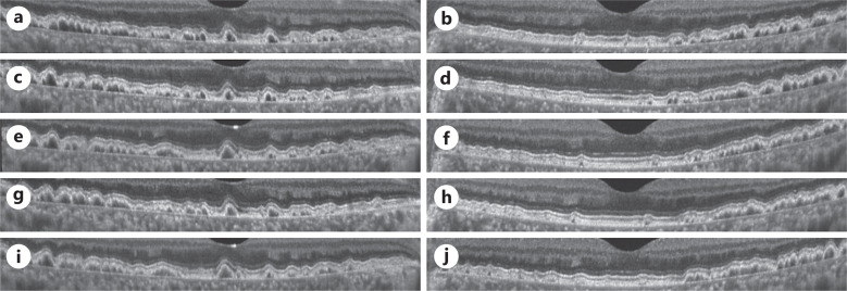
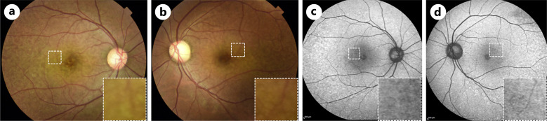
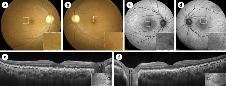
Case presentation: A 32-year-old woman was referred for ocular evaluation due to systemic steroids used for nephrotic syndrome caused by immune complex-mediated MPGN type III. She had no ocular symptoms. The initial examination showed that her best-corrected visual acuity (BCVA) was 1.0 oculus uterque (OU). Color fundus photographs revealed pigmentary abnormalities in the macular and perimacular regions OU. Fundus autofluorescence (FAF) images demonstrated numerous tiny dots with a hypoautofluorescent center surrounded by a rim of hyperautofluorescence. Enhanced depth imaging-optical coherence tomography (OCT) revealed triangular morphologic features, represented by a saw-tooth pattern with internal hyporeflectivity. Over 4.5 years, the internal reflectivity of the drusen gradually increased. Moreover, depigmentation and yellow color changes in the macular and perimacular regions worsened, without obvious changes on FAF. Her BCVA remained at 1.0 OU, without new ocular symptoms or cataract progression during the 4.5-year follow-up period. Both eyes had a significant positive correlation between swept-source OCT-based mean internal reflectivity measured by the ImageJ software and the observation period. However, no correlation was found between the estimated glomerular filtration rate (eGFR) and the observation period or between eGFR and mean internal reflectivity.
Conclusion: The internal reflectivity of cuticular drusen associated with immune complex-mediated MPGN type III showed time-dependent changes on OCT and worsened depigmentation and yellow color changes in the macular and perimacular regions, independent of renal function.
期刊介绍:
This peer-reviewed online-only journal publishes original case reports covering the entire spectrum of ophthalmology, including prevention, diagnosis, treatment, toxicities of therapy, supportive care, quality-of-life, and survivorship issues. The submission of negative results is strongly encouraged. The journal will also accept case reports dealing with the use of novel technologies, both in the arena of diagnosis and treatment. Supplementary material is welcomed. The intent of the journal is to provide clinicians and researchers with a tool to disseminate their personal experiences to a wider public as well as to review interesting cases encountered by colleagues all over the world. Universally used terms can be searched across the entire growing collection of case reports, further facilitating the retrieval of specific information. Following the open access principle, the entire contents can be retrieved at no charge, guaranteeing easy access to this valuable source of anecdotal information at all times.

 求助内容:
求助内容: 应助结果提醒方式:


