{"title":"显著性记忆:记忆提取过程中分布学习对皮质区域的影响","authors":"Cuihong Li, Jiongjiong Yang","doi":"10.1002/hbm.70301","DOIUrl":null,"url":null,"abstract":"<p>Spaced or distributed learning is an efficient way to enhance memory, especially after long retention intervals, and the lag between repetition influences memory retention. Studies have suggested that various cortical regions are involved in the spacing effect, but how the cortical regions are involved to support memory retrieval, especially at longer intervals, after DL with varying inter-study lags is still unclear. To address this issue, three groups of participants were asked to encode face–scene pairs at 20 min, 1 day, and 1 month before they were scanned by fMRI during an associative recognition task. The pairs were learned six times in three conditions: a massed (ML), distributed with a short lag (DL-S) and distributed with a long lag (DL-L). The results showed that the activation in the salience network, including the insula and cingulate cortex, was stronger when the participants retrieved the pairs correctly in the DL-L and DL-S conditions than in the ML condition. In addition, the inferior frontal gyrus/insula was more strongly activated when the new associations were correctly rejected in the DL-L than in the DL-S condition at 1 month. The functional connectivity between the hippocampus and prefrontal cortices was stronger in the DL-L than in the DL-S condition at 1 month. These results suggest that successful memory retrieval after distributed learning is associated with the regions that are responsible for salience detection and top-down control, especially at long-term retention. More salient and controlled representations could be established over time after DL and are supported by distributed brain networks.</p>","PeriodicalId":13019,"journal":{"name":"Human Brain Mapping","volume":"46 11","pages":""},"PeriodicalIF":3.3000,"publicationDate":"2025-07-31","publicationTypes":"Journal Article","fieldsOfStudy":null,"isOpenAccess":false,"openAccessPdf":"https://onlinelibrary.wiley.com/doi/epdf/10.1002/hbm.70301","citationCount":"0","resultStr":"{\"title\":\"Salient Memory: Effects of Distributed Learning on Cortical Regions During Memory Retrieval\",\"authors\":\"Cuihong Li, Jiongjiong Yang\",\"doi\":\"10.1002/hbm.70301\",\"DOIUrl\":null,\"url\":null,\"abstract\":\"<p>Spaced or distributed learning is an efficient way to enhance memory, especially after long retention intervals, and the lag between repetition influences memory retention. Studies have suggested that various cortical regions are involved in the spacing effect, but how the cortical regions are involved to support memory retrieval, especially at longer intervals, after DL with varying inter-study lags is still unclear. To address this issue, three groups of participants were asked to encode face–scene pairs at 20 min, 1 day, and 1 month before they were scanned by fMRI during an associative recognition task. The pairs were learned six times in three conditions: a massed (ML), distributed with a short lag (DL-S) and distributed with a long lag (DL-L). The results showed that the activation in the salience network, including the insula and cingulate cortex, was stronger when the participants retrieved the pairs correctly in the DL-L and DL-S conditions than in the ML condition. In addition, the inferior frontal gyrus/insula was more strongly activated when the new associations were correctly rejected in the DL-L than in the DL-S condition at 1 month. The functional connectivity between the hippocampus and prefrontal cortices was stronger in the DL-L than in the DL-S condition at 1 month. These results suggest that successful memory retrieval after distributed learning is associated with the regions that are responsible for salience detection and top-down control, especially at long-term retention. More salient and controlled representations could be established over time after DL and are supported by distributed brain networks.</p>\",\"PeriodicalId\":13019,\"journal\":{\"name\":\"Human Brain Mapping\",\"volume\":\"46 11\",\"pages\":\"\"},\"PeriodicalIF\":3.3000,\"publicationDate\":\"2025-07-31\",\"publicationTypes\":\"Journal Article\",\"fieldsOfStudy\":null,\"isOpenAccess\":false,\"openAccessPdf\":\"https://onlinelibrary.wiley.com/doi/epdf/10.1002/hbm.70301\",\"citationCount\":\"0\",\"resultStr\":null,\"platform\":\"Semanticscholar\",\"paperid\":null,\"PeriodicalName\":\"Human Brain Mapping\",\"FirstCategoryId\":\"3\",\"ListUrlMain\":\"https://onlinelibrary.wiley.com/doi/10.1002/hbm.70301\",\"RegionNum\":2,\"RegionCategory\":\"医学\",\"ArticlePicture\":[],\"TitleCN\":null,\"AbstractTextCN\":null,\"PMCID\":null,\"EPubDate\":\"\",\"PubModel\":\"\",\"JCR\":\"Q1\",\"JCRName\":\"NEUROIMAGING\",\"Score\":null,\"Total\":0}","platform":"Semanticscholar","paperid":null,"PeriodicalName":"Human Brain Mapping","FirstCategoryId":"3","ListUrlMain":"https://onlinelibrary.wiley.com/doi/10.1002/hbm.70301","RegionNum":2,"RegionCategory":"医学","ArticlePicture":[],"TitleCN":null,"AbstractTextCN":null,"PMCID":null,"EPubDate":"","PubModel":"","JCR":"Q1","JCRName":"NEUROIMAGING","Score":null,"Total":0}
Salient Memory: Effects of Distributed Learning on Cortical Regions During Memory Retrieval
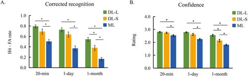
Spaced or distributed learning is an efficient way to enhance memory, especially after long retention intervals, and the lag between repetition influences memory retention. Studies have suggested that various cortical regions are involved in the spacing effect, but how the cortical regions are involved to support memory retrieval, especially at longer intervals, after DL with varying inter-study lags is still unclear. To address this issue, three groups of participants were asked to encode face–scene pairs at 20 min, 1 day, and 1 month before they were scanned by fMRI during an associative recognition task. The pairs were learned six times in three conditions: a massed (ML), distributed with a short lag (DL-S) and distributed with a long lag (DL-L). The results showed that the activation in the salience network, including the insula and cingulate cortex, was stronger when the participants retrieved the pairs correctly in the DL-L and DL-S conditions than in the ML condition. In addition, the inferior frontal gyrus/insula was more strongly activated when the new associations were correctly rejected in the DL-L than in the DL-S condition at 1 month. The functional connectivity between the hippocampus and prefrontal cortices was stronger in the DL-L than in the DL-S condition at 1 month. These results suggest that successful memory retrieval after distributed learning is associated with the regions that are responsible for salience detection and top-down control, especially at long-term retention. More salient and controlled representations could be established over time after DL and are supported by distributed brain networks.
期刊介绍:
Human Brain Mapping publishes peer-reviewed basic, clinical, technical, and theoretical research in the interdisciplinary and rapidly expanding field of human brain mapping. The journal features research derived from non-invasive brain imaging modalities used to explore the spatial and temporal organization of the neural systems supporting human behavior. Imaging modalities of interest include positron emission tomography, event-related potentials, electro-and magnetoencephalography, magnetic resonance imaging, and single-photon emission tomography. Brain mapping research in both normal and clinical populations is encouraged.
Article formats include Research Articles, Review Articles, Clinical Case Studies, and Technique, as well as Technological Developments, Theoretical Articles, and Synthetic Reviews. Technical advances, such as novel brain imaging methods, analyses for detecting or localizing neural activity, synergistic uses of multiple imaging modalities, and strategies for the design of behavioral paradigms and neural-systems modeling are of particular interest. The journal endorses the propagation of methodological standards and encourages database development in the field of human brain mapping.

 求助内容:
求助内容: 应助结果提醒方式:


