Jérémy H. Thalgott, Nicolas Zucker, Thomas Deffieux, Marit S. Koopman, Alexandre Dizeux, Cristina M. Avramut, Roman I. Koning, Hans-Jurgen Mager, Ton J. Rabelink, Mickaël Tanter, Franck Lebrin
{"title":"应用功能超声定位显微镜无创表征小鼠脑周细胞功能障碍","authors":"Jérémy H. Thalgott, Nicolas Zucker, Thomas Deffieux, Marit S. Koopman, Alexandre Dizeux, Cristina M. Avramut, Roman I. Koning, Hans-Jurgen Mager, Ton J. Rabelink, Mickaël Tanter, Franck Lebrin","doi":"10.1038/s41551-025-01465-x","DOIUrl":null,"url":null,"abstract":"<p>Early microscopic-scale pericyte dysfunction contributes to the initial stages of many neurological diseases and represents a strong candidate target for therapeutic intervention. A non-invasive imaging modality able to image microvascular alterations induced by pericyte dysfunction is needed. In addition, the development of pericyte-focused therapies remains challenging due to the lack of early biomarkers of disease progression. Here we show that cerebral microvascular alterations induced by pericyte dysfunction can be characterized non-invasively in mice using functional ultrasound localization microscopy (fULM). Depletion of endothelial endoglin in adult mice as a model of hereditary haemorrhagic telangiectasia, leads to pericyte detachment in the arteriole–capillary transition (ACT) zone. Imaging reveals that arteriolar capillaries have irregular shapes, increased diameters, reduced blood speed and neurovascular uncoupling mainly localized in the ACT zone. Transforming growth factor-β signalling activator C381 restores pericyte coverage and neurovascular response. Our study underscores the potential of fULM in characterizing early microvascular alterations. As super-resolution ultrasound transitions to the clinic, our data support its future use in monitoring pericyte-focused therapies in humans.</p>","PeriodicalId":19063,"journal":{"name":"Nature Biomedical Engineering","volume":"130 1","pages":""},"PeriodicalIF":26.8000,"publicationDate":"2025-07-30","publicationTypes":"Journal Article","fieldsOfStudy":null,"isOpenAccess":false,"openAccessPdf":"","citationCount":"0","resultStr":"{\"title\":\"Non-invasive characterization of pericyte dysfunction in mouse brain using functional ultrasound localization microscopy\",\"authors\":\"Jérémy H. Thalgott, Nicolas Zucker, Thomas Deffieux, Marit S. Koopman, Alexandre Dizeux, Cristina M. Avramut, Roman I. Koning, Hans-Jurgen Mager, Ton J. Rabelink, Mickaël Tanter, Franck Lebrin\",\"doi\":\"10.1038/s41551-025-01465-x\",\"DOIUrl\":null,\"url\":null,\"abstract\":\"<p>Early microscopic-scale pericyte dysfunction contributes to the initial stages of many neurological diseases and represents a strong candidate target for therapeutic intervention. A non-invasive imaging modality able to image microvascular alterations induced by pericyte dysfunction is needed. In addition, the development of pericyte-focused therapies remains challenging due to the lack of early biomarkers of disease progression. Here we show that cerebral microvascular alterations induced by pericyte dysfunction can be characterized non-invasively in mice using functional ultrasound localization microscopy (fULM). Depletion of endothelial endoglin in adult mice as a model of hereditary haemorrhagic telangiectasia, leads to pericyte detachment in the arteriole–capillary transition (ACT) zone. Imaging reveals that arteriolar capillaries have irregular shapes, increased diameters, reduced blood speed and neurovascular uncoupling mainly localized in the ACT zone. Transforming growth factor-β signalling activator C381 restores pericyte coverage and neurovascular response. Our study underscores the potential of fULM in characterizing early microvascular alterations. As super-resolution ultrasound transitions to the clinic, our data support its future use in monitoring pericyte-focused therapies in humans.</p>\",\"PeriodicalId\":19063,\"journal\":{\"name\":\"Nature Biomedical Engineering\",\"volume\":\"130 1\",\"pages\":\"\"},\"PeriodicalIF\":26.8000,\"publicationDate\":\"2025-07-30\",\"publicationTypes\":\"Journal Article\",\"fieldsOfStudy\":null,\"isOpenAccess\":false,\"openAccessPdf\":\"\",\"citationCount\":\"0\",\"resultStr\":null,\"platform\":\"Semanticscholar\",\"paperid\":null,\"PeriodicalName\":\"Nature Biomedical Engineering\",\"FirstCategoryId\":\"5\",\"ListUrlMain\":\"https://doi.org/10.1038/s41551-025-01465-x\",\"RegionNum\":1,\"RegionCategory\":\"医学\",\"ArticlePicture\":[],\"TitleCN\":null,\"AbstractTextCN\":null,\"PMCID\":null,\"EPubDate\":\"\",\"PubModel\":\"\",\"JCR\":\"Q1\",\"JCRName\":\"ENGINEERING, BIOMEDICAL\",\"Score\":null,\"Total\":0}","platform":"Semanticscholar","paperid":null,"PeriodicalName":"Nature Biomedical Engineering","FirstCategoryId":"5","ListUrlMain":"https://doi.org/10.1038/s41551-025-01465-x","RegionNum":1,"RegionCategory":"医学","ArticlePicture":[],"TitleCN":null,"AbstractTextCN":null,"PMCID":null,"EPubDate":"","PubModel":"","JCR":"Q1","JCRName":"ENGINEERING, BIOMEDICAL","Score":null,"Total":0}
Non-invasive characterization of pericyte dysfunction in mouse brain using functional ultrasound localization microscopy
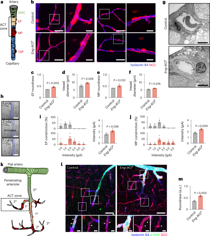
Early microscopic-scale pericyte dysfunction contributes to the initial stages of many neurological diseases and represents a strong candidate target for therapeutic intervention. A non-invasive imaging modality able to image microvascular alterations induced by pericyte dysfunction is needed. In addition, the development of pericyte-focused therapies remains challenging due to the lack of early biomarkers of disease progression. Here we show that cerebral microvascular alterations induced by pericyte dysfunction can be characterized non-invasively in mice using functional ultrasound localization microscopy (fULM). Depletion of endothelial endoglin in adult mice as a model of hereditary haemorrhagic telangiectasia, leads to pericyte detachment in the arteriole–capillary transition (ACT) zone. Imaging reveals that arteriolar capillaries have irregular shapes, increased diameters, reduced blood speed and neurovascular uncoupling mainly localized in the ACT zone. Transforming growth factor-β signalling activator C381 restores pericyte coverage and neurovascular response. Our study underscores the potential of fULM in characterizing early microvascular alterations. As super-resolution ultrasound transitions to the clinic, our data support its future use in monitoring pericyte-focused therapies in humans.
期刊介绍:
Nature Biomedical Engineering is an online-only monthly journal that was launched in January 2017. It aims to publish original research, reviews, and commentary focusing on applied biomedicine and health technology. The journal targets a diverse audience, including life scientists who are involved in developing experimental or computational systems and methods to enhance our understanding of human physiology. It also covers biomedical researchers and engineers who are engaged in designing or optimizing therapies, assays, devices, or procedures for diagnosing or treating diseases. Additionally, clinicians, who make use of research outputs to evaluate patient health or administer therapy in various clinical settings and healthcare contexts, are also part of the target audience.
 求助内容:
求助内容: 应助结果提醒方式:


