Kento Ishikawa, James K Chambers, Ko Nakashima, Takehiro Sakai, Kazuyuki Uchida
{"title":"幼猫肉芽肿性结肠炎伴牛原鞘感染。","authors":"Kento Ishikawa, James K Chambers, Ko Nakashima, Takehiro Sakai, Kazuyuki Uchida","doi":"10.1177/20551169251348548","DOIUrl":null,"url":null,"abstract":"<p><strong>Case summary: </strong>A 1-year-old spayed female mixed-breed cat presented with chronic large bowel diarrhoea. The cat was treated with various antibiotics, prednisolone and dietary supplements without significant improvement. Endoscopic examination revealed an irregular colonic mucosa with multifocal erosion and haemorrhage. Impression smears of the colonic mucosa contained large numbers of unicellular round-to-oval organisms and macrophages. Histologically, granulomatous inflammation with numerous organisms was observed in the lamina propria of the colonic mucosa. The organisms had a cell wall and internal segmentation, which were positive on periodic acid-Schiff and Grocott's methenamine silver staining. Immunohistochemically, CD204-positive macrophages had accumulated in the lesions. Ultrastructural examination revealed dense bodies and starch granules, and the absence of chloroplasts in the cytoplasm of the organisms. PCR and a sequence analysis detected the <i>Prototheca bovis 18S rRNA</i> gene in formalin-fixed, paraffin-embedded colonic mucosa tissue. Based on these findings, the cat was diagnosed with granulomatous colitis associated with <i>P bovis</i> infection.</p><p><strong>Relevance and novel information: </strong>This first report of feline intestinal protothecosis suggests that <i>Prototheca</i> species infection should be considered in the differential diagnosis of cats with treatment-resistant chronic large bowel diarrhoea.</p>","PeriodicalId":36588,"journal":{"name":"Journal of Feline Medicine and Surgery Open Reports","volume":"11 2","pages":"20551169251348548"},"PeriodicalIF":0.7000,"publicationDate":"2025-07-22","publicationTypes":"Journal Article","fieldsOfStudy":null,"isOpenAccess":false,"openAccessPdf":"https://www.ncbi.nlm.nih.gov/pmc/articles/PMC12290248/pdf/","citationCount":"0","resultStr":"{\"title\":\"Granulomatous colitis associated with <i>Prototheca bovis</i> infection in a young cat.\",\"authors\":\"Kento Ishikawa, James K Chambers, Ko Nakashima, Takehiro Sakai, Kazuyuki Uchida\",\"doi\":\"10.1177/20551169251348548\",\"DOIUrl\":null,\"url\":null,\"abstract\":\"<p><strong>Case summary: </strong>A 1-year-old spayed female mixed-breed cat presented with chronic large bowel diarrhoea. The cat was treated with various antibiotics, prednisolone and dietary supplements without significant improvement. Endoscopic examination revealed an irregular colonic mucosa with multifocal erosion and haemorrhage. Impression smears of the colonic mucosa contained large numbers of unicellular round-to-oval organisms and macrophages. Histologically, granulomatous inflammation with numerous organisms was observed in the lamina propria of the colonic mucosa. The organisms had a cell wall and internal segmentation, which were positive on periodic acid-Schiff and Grocott's methenamine silver staining. Immunohistochemically, CD204-positive macrophages had accumulated in the lesions. Ultrastructural examination revealed dense bodies and starch granules, and the absence of chloroplasts in the cytoplasm of the organisms. PCR and a sequence analysis detected the <i>Prototheca bovis 18S rRNA</i> gene in formalin-fixed, paraffin-embedded colonic mucosa tissue. Based on these findings, the cat was diagnosed with granulomatous colitis associated with <i>P bovis</i> infection.</p><p><strong>Relevance and novel information: </strong>This first report of feline intestinal protothecosis suggests that <i>Prototheca</i> species infection should be considered in the differential diagnosis of cats with treatment-resistant chronic large bowel diarrhoea.</p>\",\"PeriodicalId\":36588,\"journal\":{\"name\":\"Journal of Feline Medicine and Surgery Open Reports\",\"volume\":\"11 2\",\"pages\":\"20551169251348548\"},\"PeriodicalIF\":0.7000,\"publicationDate\":\"2025-07-22\",\"publicationTypes\":\"Journal Article\",\"fieldsOfStudy\":null,\"isOpenAccess\":false,\"openAccessPdf\":\"https://www.ncbi.nlm.nih.gov/pmc/articles/PMC12290248/pdf/\",\"citationCount\":\"0\",\"resultStr\":null,\"platform\":\"Semanticscholar\",\"paperid\":null,\"PeriodicalName\":\"Journal of Feline Medicine and Surgery Open Reports\",\"FirstCategoryId\":\"1085\",\"ListUrlMain\":\"https://doi.org/10.1177/20551169251348548\",\"RegionNum\":0,\"RegionCategory\":null,\"ArticlePicture\":[],\"TitleCN\":null,\"AbstractTextCN\":null,\"PMCID\":null,\"EPubDate\":\"2025/7/1 0:00:00\",\"PubModel\":\"eCollection\",\"JCR\":\"Q3\",\"JCRName\":\"VETERINARY SCIENCES\",\"Score\":null,\"Total\":0}","platform":"Semanticscholar","paperid":null,"PeriodicalName":"Journal of Feline Medicine and Surgery Open Reports","FirstCategoryId":"1085","ListUrlMain":"https://doi.org/10.1177/20551169251348548","RegionNum":0,"RegionCategory":null,"ArticlePicture":[],"TitleCN":null,"AbstractTextCN":null,"PMCID":null,"EPubDate":"2025/7/1 0:00:00","PubModel":"eCollection","JCR":"Q3","JCRName":"VETERINARY SCIENCES","Score":null,"Total":0}
Granulomatous colitis associated with Prototheca bovis infection in a young cat.
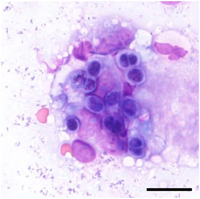
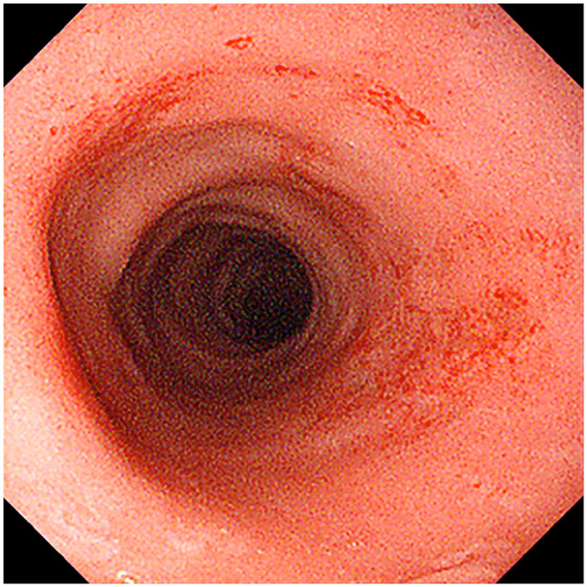
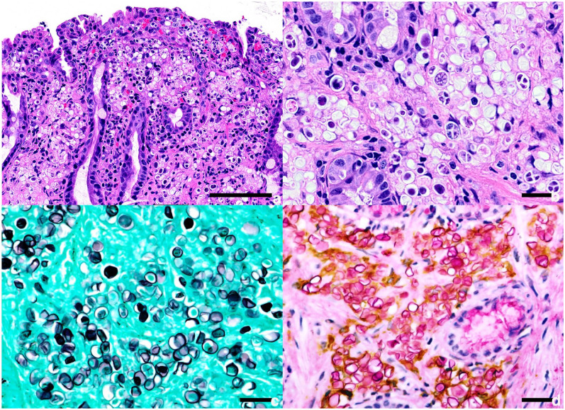
Case summary: A 1-year-old spayed female mixed-breed cat presented with chronic large bowel diarrhoea. The cat was treated with various antibiotics, prednisolone and dietary supplements without significant improvement. Endoscopic examination revealed an irregular colonic mucosa with multifocal erosion and haemorrhage. Impression smears of the colonic mucosa contained large numbers of unicellular round-to-oval organisms and macrophages. Histologically, granulomatous inflammation with numerous organisms was observed in the lamina propria of the colonic mucosa. The organisms had a cell wall and internal segmentation, which were positive on periodic acid-Schiff and Grocott's methenamine silver staining. Immunohistochemically, CD204-positive macrophages had accumulated in the lesions. Ultrastructural examination revealed dense bodies and starch granules, and the absence of chloroplasts in the cytoplasm of the organisms. PCR and a sequence analysis detected the Prototheca bovis 18S rRNA gene in formalin-fixed, paraffin-embedded colonic mucosa tissue. Based on these findings, the cat was diagnosed with granulomatous colitis associated with P bovis infection.
Relevance and novel information: This first report of feline intestinal protothecosis suggests that Prototheca species infection should be considered in the differential diagnosis of cats with treatment-resistant chronic large bowel diarrhoea.

 求助内容:
求助内容: 应助结果提醒方式:


