Camila Soares Franco, Paula Terra Martins Almeida Amaral, Eduardo Kaiser Ururahy Nunes Fonseca, Tassia Regina Yamanari, Paulo Esrom Moreira Catarina, Alessandra de Pinho Pimenta Borges, Luiz Augusto de Moraes Pinheiro Filho, Felipe Freitas Camara, Márcio Valente Yamada Sawamura
{"title":"286例胸部计算机断层反转晕征回顾性分析。","authors":"Camila Soares Franco, Paula Terra Martins Almeida Amaral, Eduardo Kaiser Ururahy Nunes Fonseca, Tassia Regina Yamanari, Paulo Esrom Moreira Catarina, Alessandra de Pinho Pimenta Borges, Luiz Augusto de Moraes Pinheiro Filho, Felipe Freitas Camara, Márcio Valente Yamada Sawamura","doi":"10.1590/0100-3984.2025.0014-en","DOIUrl":null,"url":null,"abstract":"<p><strong>Objective: </strong>To characterize the main causes of the reversed halo sign (RHS) on computed tomography (CT) of the chest and its imaging features.</p><p><strong>Materials and methods: </strong>This was a retrospective study reviewing all chest CT scans for which the report contained the term \"reversed halo sign\" among those performed between 2015 and 2020 at a tertiary care hospital.</p><p><strong>Results: </strong>A total of 286 cases were identified, and the corresponding CT images and clinical data were reviewed. In this population, the most common cause of an RHS was pulmonary infarction (in 42%), followed by cryptogenic organizing pneumonia (in 17%) and bacterial pneumonia (in 16%). In addition, the CT characteristics of the RHS were identified in various conditions, such as pulmonary thromboembolism with pulmonary infarction, in which the RHS was typically smooth-walled and solitary with a peripheral distribution.</p><p><strong>Conclusion: </strong>The RHS can be observed in many contexts, and its CT characteristics, in combination with the clinical picture, can help narrow the differential diagnosis.</p>","PeriodicalId":20842,"journal":{"name":"Radiologia Brasileira","volume":"58 ","pages":"e20250014"},"PeriodicalIF":0.0000,"publicationDate":"2025-07-17","publicationTypes":"Journal Article","fieldsOfStudy":null,"isOpenAccess":false,"openAccessPdf":"https://www.ncbi.nlm.nih.gov/pmc/articles/PMC12295819/pdf/","citationCount":"0","resultStr":"{\"title\":\"Reversed halo sign on chest computed tomography: a retrospective analysis of 286 cases.\",\"authors\":\"Camila Soares Franco, Paula Terra Martins Almeida Amaral, Eduardo Kaiser Ururahy Nunes Fonseca, Tassia Regina Yamanari, Paulo Esrom Moreira Catarina, Alessandra de Pinho Pimenta Borges, Luiz Augusto de Moraes Pinheiro Filho, Felipe Freitas Camara, Márcio Valente Yamada Sawamura\",\"doi\":\"10.1590/0100-3984.2025.0014-en\",\"DOIUrl\":null,\"url\":null,\"abstract\":\"<p><strong>Objective: </strong>To characterize the main causes of the reversed halo sign (RHS) on computed tomography (CT) of the chest and its imaging features.</p><p><strong>Materials and methods: </strong>This was a retrospective study reviewing all chest CT scans for which the report contained the term \\\"reversed halo sign\\\" among those performed between 2015 and 2020 at a tertiary care hospital.</p><p><strong>Results: </strong>A total of 286 cases were identified, and the corresponding CT images and clinical data were reviewed. In this population, the most common cause of an RHS was pulmonary infarction (in 42%), followed by cryptogenic organizing pneumonia (in 17%) and bacterial pneumonia (in 16%). In addition, the CT characteristics of the RHS were identified in various conditions, such as pulmonary thromboembolism with pulmonary infarction, in which the RHS was typically smooth-walled and solitary with a peripheral distribution.</p><p><strong>Conclusion: </strong>The RHS can be observed in many contexts, and its CT characteristics, in combination with the clinical picture, can help narrow the differential diagnosis.</p>\",\"PeriodicalId\":20842,\"journal\":{\"name\":\"Radiologia Brasileira\",\"volume\":\"58 \",\"pages\":\"e20250014\"},\"PeriodicalIF\":0.0000,\"publicationDate\":\"2025-07-17\",\"publicationTypes\":\"Journal Article\",\"fieldsOfStudy\":null,\"isOpenAccess\":false,\"openAccessPdf\":\"https://www.ncbi.nlm.nih.gov/pmc/articles/PMC12295819/pdf/\",\"citationCount\":\"0\",\"resultStr\":null,\"platform\":\"Semanticscholar\",\"paperid\":null,\"PeriodicalName\":\"Radiologia Brasileira\",\"FirstCategoryId\":\"1085\",\"ListUrlMain\":\"https://doi.org/10.1590/0100-3984.2025.0014-en\",\"RegionNum\":0,\"RegionCategory\":null,\"ArticlePicture\":[],\"TitleCN\":null,\"AbstractTextCN\":null,\"PMCID\":null,\"EPubDate\":\"2025/1/1 0:00:00\",\"PubModel\":\"eCollection\",\"JCR\":\"Q3\",\"JCRName\":\"Medicine\",\"Score\":null,\"Total\":0}","platform":"Semanticscholar","paperid":null,"PeriodicalName":"Radiologia Brasileira","FirstCategoryId":"1085","ListUrlMain":"https://doi.org/10.1590/0100-3984.2025.0014-en","RegionNum":0,"RegionCategory":null,"ArticlePicture":[],"TitleCN":null,"AbstractTextCN":null,"PMCID":null,"EPubDate":"2025/1/1 0:00:00","PubModel":"eCollection","JCR":"Q3","JCRName":"Medicine","Score":null,"Total":0}
Reversed halo sign on chest computed tomography: a retrospective analysis of 286 cases.
Objective: To characterize the main causes of the reversed halo sign (RHS) on computed tomography (CT) of the chest and its imaging features.
Materials and methods: This was a retrospective study reviewing all chest CT scans for which the report contained the term "reversed halo sign" among those performed between 2015 and 2020 at a tertiary care hospital.
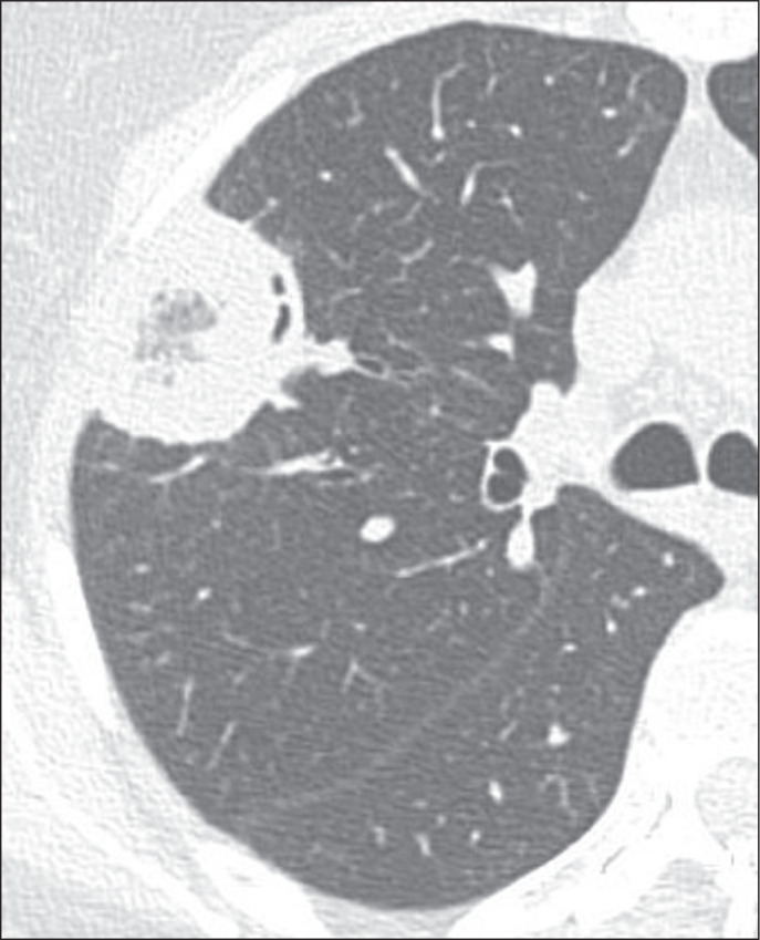
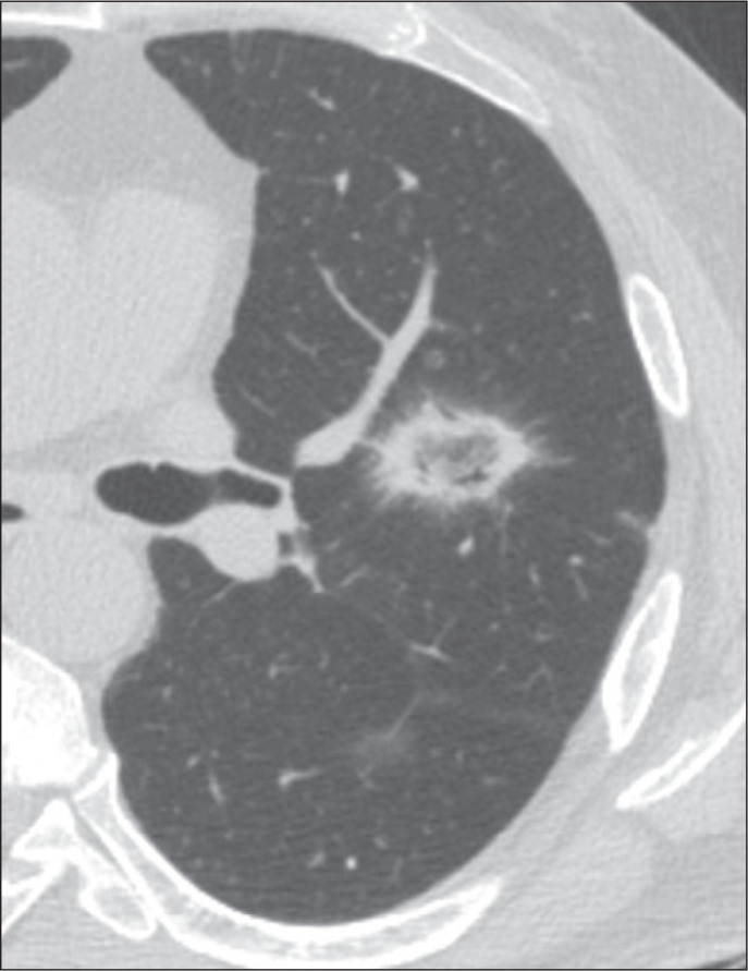
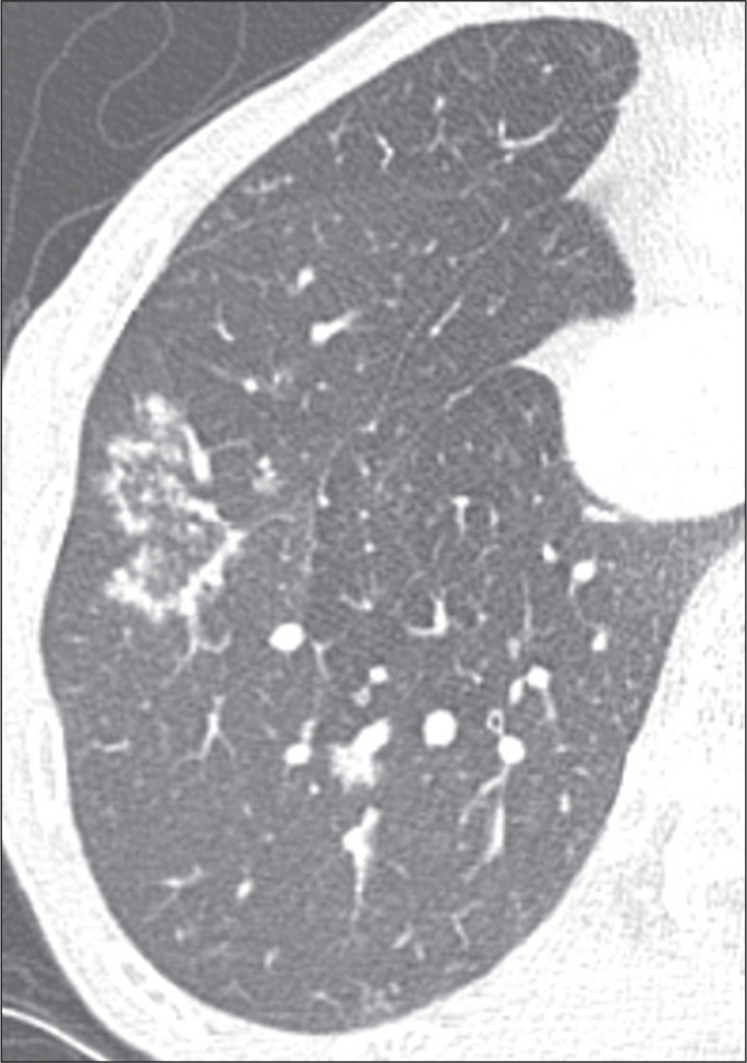
Results: A total of 286 cases were identified, and the corresponding CT images and clinical data were reviewed. In this population, the most common cause of an RHS was pulmonary infarction (in 42%), followed by cryptogenic organizing pneumonia (in 17%) and bacterial pneumonia (in 16%). In addition, the CT characteristics of the RHS were identified in various conditions, such as pulmonary thromboembolism with pulmonary infarction, in which the RHS was typically smooth-walled and solitary with a peripheral distribution.
Conclusion: The RHS can be observed in many contexts, and its CT characteristics, in combination with the clinical picture, can help narrow the differential diagnosis.

 求助内容:
求助内容: 应助结果提醒方式:


