{"title":"利用微计算机断层扫描技术对光纤中子探测器中一小片掺铈锂玻璃闪烁体进行三维重建。","authors":"Akihisa Ishikawa, Mariko Segawa, Yosuke Toh, Kenichi Watanabe, Akihiko Masuda, Tetsuro Matsumoto, Atsushi Yamazaki, Sachiko Yoshihashi, Akira Uritani, Hideki Harano","doi":"10.1093/jrr/rraf048","DOIUrl":null,"url":null,"abstract":"<p><p>An optical fiber-based neutron detector is a real-time neutron monitor for an intense neutron field. A small piece of neutron scintillator, such as Ce-doped lithium glass (Li-glass), used in the detector has a random shape with a grain size of 200-400 μm. This causes shape-dependent effects on the detector response. However, it is difficult to control or determine its shape due to its small size. Here we propose a technique to characterize the fine structure of a small piece of scintillator using a microcomputed tomography (CT) system. To verify accuracy, the mass calculated based on the density of Li-glass and the volume extracted from the obtained CT image was compared to the mass measured in advance using an electronic balance. In the obtained CT images, the fine shape of the small piece of Li-glass was clearly visible, and no false signals from the surrounding components were observed. The calculated mass was in good agreement with the measured value, however, when the total number of projection images was 2000, a slight underestimation was observed. This was mitigated by increasing the number of projection images, and the difference between the calculated and measured mass was 1.6% when the number of the projection images was 3141. This was equivalent to the uncertainty of the measured mass. The proposed technique will be useful when high accuracy is needed, such as for medical applications.</p>","PeriodicalId":16922,"journal":{"name":"Journal of Radiation Research","volume":" ","pages":"563-569"},"PeriodicalIF":2.0000,"publicationDate":"2025-09-23","publicationTypes":"Journal Article","fieldsOfStudy":null,"isOpenAccess":false,"openAccessPdf":"https://www.ncbi.nlm.nih.gov/pmc/articles/PMC12460048/pdf/","citationCount":"0","resultStr":"{\"title\":\"Three-dimensional reconstruction of a small piece of Ce-doped lithium glass scintillator of an optical fiber-based neutron detector using microcomputed tomography technique.\",\"authors\":\"Akihisa Ishikawa, Mariko Segawa, Yosuke Toh, Kenichi Watanabe, Akihiko Masuda, Tetsuro Matsumoto, Atsushi Yamazaki, Sachiko Yoshihashi, Akira Uritani, Hideki Harano\",\"doi\":\"10.1093/jrr/rraf048\",\"DOIUrl\":null,\"url\":null,\"abstract\":\"<p><p>An optical fiber-based neutron detector is a real-time neutron monitor for an intense neutron field. A small piece of neutron scintillator, such as Ce-doped lithium glass (Li-glass), used in the detector has a random shape with a grain size of 200-400 μm. This causes shape-dependent effects on the detector response. However, it is difficult to control or determine its shape due to its small size. Here we propose a technique to characterize the fine structure of a small piece of scintillator using a microcomputed tomography (CT) system. To verify accuracy, the mass calculated based on the density of Li-glass and the volume extracted from the obtained CT image was compared to the mass measured in advance using an electronic balance. In the obtained CT images, the fine shape of the small piece of Li-glass was clearly visible, and no false signals from the surrounding components were observed. The calculated mass was in good agreement with the measured value, however, when the total number of projection images was 2000, a slight underestimation was observed. This was mitigated by increasing the number of projection images, and the difference between the calculated and measured mass was 1.6% when the number of the projection images was 3141. This was equivalent to the uncertainty of the measured mass. The proposed technique will be useful when high accuracy is needed, such as for medical applications.</p>\",\"PeriodicalId\":16922,\"journal\":{\"name\":\"Journal of Radiation Research\",\"volume\":\" \",\"pages\":\"563-569\"},\"PeriodicalIF\":2.0000,\"publicationDate\":\"2025-09-23\",\"publicationTypes\":\"Journal Article\",\"fieldsOfStudy\":null,\"isOpenAccess\":false,\"openAccessPdf\":\"https://www.ncbi.nlm.nih.gov/pmc/articles/PMC12460048/pdf/\",\"citationCount\":\"0\",\"resultStr\":null,\"platform\":\"Semanticscholar\",\"paperid\":null,\"PeriodicalName\":\"Journal of Radiation Research\",\"FirstCategoryId\":\"3\",\"ListUrlMain\":\"https://doi.org/10.1093/jrr/rraf048\",\"RegionNum\":4,\"RegionCategory\":\"医学\",\"ArticlePicture\":[],\"TitleCN\":null,\"AbstractTextCN\":null,\"PMCID\":null,\"EPubDate\":\"\",\"PubModel\":\"\",\"JCR\":\"Q2\",\"JCRName\":\"BIOLOGY\",\"Score\":null,\"Total\":0}","platform":"Semanticscholar","paperid":null,"PeriodicalName":"Journal of Radiation Research","FirstCategoryId":"3","ListUrlMain":"https://doi.org/10.1093/jrr/rraf048","RegionNum":4,"RegionCategory":"医学","ArticlePicture":[],"TitleCN":null,"AbstractTextCN":null,"PMCID":null,"EPubDate":"","PubModel":"","JCR":"Q2","JCRName":"BIOLOGY","Score":null,"Total":0}
Three-dimensional reconstruction of a small piece of Ce-doped lithium glass scintillator of an optical fiber-based neutron detector using microcomputed tomography technique.
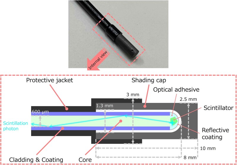
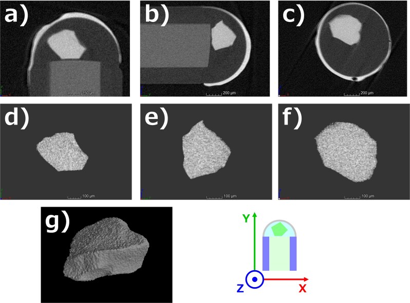
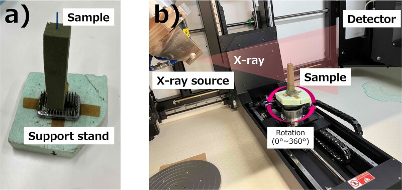
An optical fiber-based neutron detector is a real-time neutron monitor for an intense neutron field. A small piece of neutron scintillator, such as Ce-doped lithium glass (Li-glass), used in the detector has a random shape with a grain size of 200-400 μm. This causes shape-dependent effects on the detector response. However, it is difficult to control or determine its shape due to its small size. Here we propose a technique to characterize the fine structure of a small piece of scintillator using a microcomputed tomography (CT) system. To verify accuracy, the mass calculated based on the density of Li-glass and the volume extracted from the obtained CT image was compared to the mass measured in advance using an electronic balance. In the obtained CT images, the fine shape of the small piece of Li-glass was clearly visible, and no false signals from the surrounding components were observed. The calculated mass was in good agreement with the measured value, however, when the total number of projection images was 2000, a slight underestimation was observed. This was mitigated by increasing the number of projection images, and the difference between the calculated and measured mass was 1.6% when the number of the projection images was 3141. This was equivalent to the uncertainty of the measured mass. The proposed technique will be useful when high accuracy is needed, such as for medical applications.
期刊介绍:
The Journal of Radiation Research (JRR) is an official journal of The Japanese Radiation Research Society (JRRS), and the Japanese Society for Radiation Oncology (JASTRO).
Since its launch in 1960 as the official journal of the JRRS, the journal has published scientific articles in radiation science in biology, chemistry, physics, epidemiology, and environmental sciences. JRR broadened its scope to include oncology in 2009, when JASTRO partnered with the JRRS to publish the journal.
Articles considered fall into two broad categories:
Oncology & Medicine - including all aspects of research with patients that impacts on the treatment of cancer using radiation. Papers which cover related radiation therapies, radiation dosimetry, and those describing the basis for treatment methods including techniques, are also welcomed. Clinical case reports are not acceptable.
Radiation Research - basic science studies of radiation effects on livings in the area of physics, chemistry, biology, epidemiology and environmental sciences.
Please be advised that JRR does not accept any papers of pure physics or chemistry.
The journal is bimonthly, and is edited and published by the JRR Editorial Committee.

 求助内容:
求助内容: 应助结果提醒方式:


