Liubov A Tashireva, Anna Yu Kalinchuk, Elena O Shmakova, Elisaveta A Tsarenkova, Dmitriy M Loos, Pavel Iamschikov, Ivan A Patskan, Alexandra V Avgustinovich, Sergey V Vtorushin, Irina V Larionova, Evgeniya S Grigorieva
{"title":"肿瘤微环境中pd -1阳性CD8+ T细胞和pd -1阳性FoxP3+细胞预测胃癌患者对新辅助化疗免疫治疗的反应","authors":"Liubov A Tashireva, Anna Yu Kalinchuk, Elena O Shmakova, Elisaveta A Tsarenkova, Dmitriy M Loos, Pavel Iamschikov, Ivan A Patskan, Alexandra V Avgustinovich, Sergey V Vtorushin, Irina V Larionova, Evgeniya S Grigorieva","doi":"10.3390/cancers17142407","DOIUrl":null,"url":null,"abstract":"<p><strong>Background/objectives: </strong>In gastric cancer, only a subset of patients benefit clinically from neoadjuvant chemoimmunotherapy, underscoring the need for robust biomarkers that can predict treatment responses and guide personalized immunotherapy. This study aimed to characterize the immune microenvironment of gastric tumors and identify predictive markers associated with therapeutic efficacy.</p><p><strong>Methods: </strong>We prospectively enrolled 16 patients with histologically confirmed, PD-L1-positive (CPS ≥ 1) gastric adenocarcinoma (T<sub>2-4</sub>N<sub>0-1</sub>M<sub>0</sub>). All patients received eight cycles of FLOT chemotherapy combined with pembrolizumab. Treatment response was assessed by Mandard tumor regression grading. Spatial transcriptomic profiling (10x Genomics Visium) and multiplex immunofluorescence were used to evaluate tumor-infiltrating immune cell subsets and PD-1 expression at baseline and after treatment.</p><p><strong>Results: </strong>Transcriptomic analysis differentiated the immune landscapes of responders from non-responders. Responders exhibited elevated expression of <i>IL1B</i>, <i>CXCL5</i>, <i>HMGB1</i>, and <i>IFNGR2</i>, indicative of an inflamed tumor microenvironment and type I/II interferon signaling. In contrast, non-responders demonstrated upregulation of immunosuppressive genes such as <i>LGALS3</i>, <i>IDO1</i>, and <i>CD55</i>, along with enrichment in oxidative phosphorylation and antigen presentation pathways. Multiplex immunofluorescence confirmed a higher density of FoxP3<sup>+</sup> regulatory T cells in non-responders (median 5.36% vs. 2.41%; <i>p</i> = 0.0032). Notably, PD-1<sup>+</sup> CD8<sup>+</sup> T cell and PD-1<sup>+</sup> FoxP3<sup>+</sup> Treg frequencies were significantly elevated in non-responders, suggesting that PD-1 expression within cytotoxic and regulatory compartments may contribute to immune evasion. No substantial differences were observed in PD-L1 CPS or PD-1<sup>+</sup> B cells and PD-1<sup>+</sup> macrophages.</p><p><strong>Conclusions: </strong>Our findings identify PD-1<sup>+</sup> CD8<sup>+</sup> T cells and PD-1<sup>+</sup> FoxP3<sup>+</sup> Tregs as potential biomarkers of resistance to neoadjuvant chemoimmunotherapy in gastric cancer. Transcriptional programs centered on IL1B/CXCL5 and LGALS3/IDO1 define distinct immune phenotypes that may guide future combination strategies targeting both effector and suppressive arms of the tumor immune response.</p>","PeriodicalId":9681,"journal":{"name":"Cancers","volume":"17 14","pages":""},"PeriodicalIF":4.4000,"publicationDate":"2025-07-21","publicationTypes":"Journal Article","fieldsOfStudy":null,"isOpenAccess":false,"openAccessPdf":"https://www.ncbi.nlm.nih.gov/pmc/articles/PMC12293087/pdf/","citationCount":"0","resultStr":"{\"title\":\"PD-1-Positive CD8+ T Cells and PD-1-Positive FoxP3+ Cells in Tumor Microenvironment Predict Response to Neoadjuvant Chemoimmunotherapy in Gastric Cancer Patients.\",\"authors\":\"Liubov A Tashireva, Anna Yu Kalinchuk, Elena O Shmakova, Elisaveta A Tsarenkova, Dmitriy M Loos, Pavel Iamschikov, Ivan A Patskan, Alexandra V Avgustinovich, Sergey V Vtorushin, Irina V Larionova, Evgeniya S Grigorieva\",\"doi\":\"10.3390/cancers17142407\",\"DOIUrl\":null,\"url\":null,\"abstract\":\"<p><strong>Background/objectives: </strong>In gastric cancer, only a subset of patients benefit clinically from neoadjuvant chemoimmunotherapy, underscoring the need for robust biomarkers that can predict treatment responses and guide personalized immunotherapy. This study aimed to characterize the immune microenvironment of gastric tumors and identify predictive markers associated with therapeutic efficacy.</p><p><strong>Methods: </strong>We prospectively enrolled 16 patients with histologically confirmed, PD-L1-positive (CPS ≥ 1) gastric adenocarcinoma (T<sub>2-4</sub>N<sub>0-1</sub>M<sub>0</sub>). All patients received eight cycles of FLOT chemotherapy combined with pembrolizumab. Treatment response was assessed by Mandard tumor regression grading. Spatial transcriptomic profiling (10x Genomics Visium) and multiplex immunofluorescence were used to evaluate tumor-infiltrating immune cell subsets and PD-1 expression at baseline and after treatment.</p><p><strong>Results: </strong>Transcriptomic analysis differentiated the immune landscapes of responders from non-responders. Responders exhibited elevated expression of <i>IL1B</i>, <i>CXCL5</i>, <i>HMGB1</i>, and <i>IFNGR2</i>, indicative of an inflamed tumor microenvironment and type I/II interferon signaling. In contrast, non-responders demonstrated upregulation of immunosuppressive genes such as <i>LGALS3</i>, <i>IDO1</i>, and <i>CD55</i>, along with enrichment in oxidative phosphorylation and antigen presentation pathways. Multiplex immunofluorescence confirmed a higher density of FoxP3<sup>+</sup> regulatory T cells in non-responders (median 5.36% vs. 2.41%; <i>p</i> = 0.0032). Notably, PD-1<sup>+</sup> CD8<sup>+</sup> T cell and PD-1<sup>+</sup> FoxP3<sup>+</sup> Treg frequencies were significantly elevated in non-responders, suggesting that PD-1 expression within cytotoxic and regulatory compartments may contribute to immune evasion. No substantial differences were observed in PD-L1 CPS or PD-1<sup>+</sup> B cells and PD-1<sup>+</sup> macrophages.</p><p><strong>Conclusions: </strong>Our findings identify PD-1<sup>+</sup> CD8<sup>+</sup> T cells and PD-1<sup>+</sup> FoxP3<sup>+</sup> Tregs as potential biomarkers of resistance to neoadjuvant chemoimmunotherapy in gastric cancer. Transcriptional programs centered on IL1B/CXCL5 and LGALS3/IDO1 define distinct immune phenotypes that may guide future combination strategies targeting both effector and suppressive arms of the tumor immune response.</p>\",\"PeriodicalId\":9681,\"journal\":{\"name\":\"Cancers\",\"volume\":\"17 14\",\"pages\":\"\"},\"PeriodicalIF\":4.4000,\"publicationDate\":\"2025-07-21\",\"publicationTypes\":\"Journal Article\",\"fieldsOfStudy\":null,\"isOpenAccess\":false,\"openAccessPdf\":\"https://www.ncbi.nlm.nih.gov/pmc/articles/PMC12293087/pdf/\",\"citationCount\":\"0\",\"resultStr\":null,\"platform\":\"Semanticscholar\",\"paperid\":null,\"PeriodicalName\":\"Cancers\",\"FirstCategoryId\":\"3\",\"ListUrlMain\":\"https://doi.org/10.3390/cancers17142407\",\"RegionNum\":2,\"RegionCategory\":\"医学\",\"ArticlePicture\":[],\"TitleCN\":null,\"AbstractTextCN\":null,\"PMCID\":null,\"EPubDate\":\"\",\"PubModel\":\"\",\"JCR\":\"Q1\",\"JCRName\":\"ONCOLOGY\",\"Score\":null,\"Total\":0}","platform":"Semanticscholar","paperid":null,"PeriodicalName":"Cancers","FirstCategoryId":"3","ListUrlMain":"https://doi.org/10.3390/cancers17142407","RegionNum":2,"RegionCategory":"医学","ArticlePicture":[],"TitleCN":null,"AbstractTextCN":null,"PMCID":null,"EPubDate":"","PubModel":"","JCR":"Q1","JCRName":"ONCOLOGY","Score":null,"Total":0}
PD-1-Positive CD8+ T Cells and PD-1-Positive FoxP3+ Cells in Tumor Microenvironment Predict Response to Neoadjuvant Chemoimmunotherapy in Gastric Cancer Patients.
Background/objectives: In gastric cancer, only a subset of patients benefit clinically from neoadjuvant chemoimmunotherapy, underscoring the need for robust biomarkers that can predict treatment responses and guide personalized immunotherapy. This study aimed to characterize the immune microenvironment of gastric tumors and identify predictive markers associated with therapeutic efficacy.
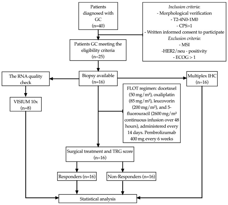
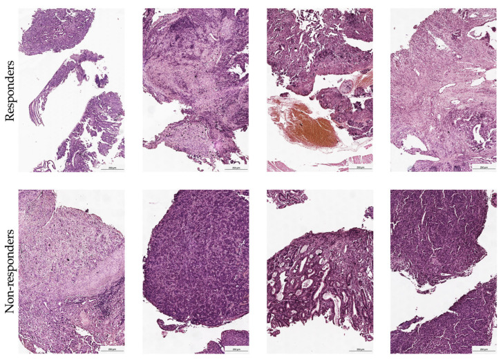
Methods: We prospectively enrolled 16 patients with histologically confirmed, PD-L1-positive (CPS ≥ 1) gastric adenocarcinoma (T2-4N0-1M0). All patients received eight cycles of FLOT chemotherapy combined with pembrolizumab. Treatment response was assessed by Mandard tumor regression grading. Spatial transcriptomic profiling (10x Genomics Visium) and multiplex immunofluorescence were used to evaluate tumor-infiltrating immune cell subsets and PD-1 expression at baseline and after treatment.
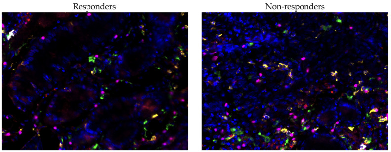
Results: Transcriptomic analysis differentiated the immune landscapes of responders from non-responders. Responders exhibited elevated expression of IL1B, CXCL5, HMGB1, and IFNGR2, indicative of an inflamed tumor microenvironment and type I/II interferon signaling. In contrast, non-responders demonstrated upregulation of immunosuppressive genes such as LGALS3, IDO1, and CD55, along with enrichment in oxidative phosphorylation and antigen presentation pathways. Multiplex immunofluorescence confirmed a higher density of FoxP3+ regulatory T cells in non-responders (median 5.36% vs. 2.41%; p = 0.0032). Notably, PD-1+ CD8+ T cell and PD-1+ FoxP3+ Treg frequencies were significantly elevated in non-responders, suggesting that PD-1 expression within cytotoxic and regulatory compartments may contribute to immune evasion. No substantial differences were observed in PD-L1 CPS or PD-1+ B cells and PD-1+ macrophages.
Conclusions: Our findings identify PD-1+ CD8+ T cells and PD-1+ FoxP3+ Tregs as potential biomarkers of resistance to neoadjuvant chemoimmunotherapy in gastric cancer. Transcriptional programs centered on IL1B/CXCL5 and LGALS3/IDO1 define distinct immune phenotypes that may guide future combination strategies targeting both effector and suppressive arms of the tumor immune response.
期刊介绍:
Cancers (ISSN 2072-6694) is an international, peer-reviewed open access journal on oncology. It publishes reviews, regular research papers and short communications. Our aim is to encourage scientists to publish their experimental and theoretical results in as much detail as possible. There is no restriction on the length of the papers. The full experimental details must be provided so that the results can be reproduced.
 求助内容:
求助内容: 应助结果提醒方式:


