Annelise G Pageau, Lauren A Burt, Leigh Gabel, Steven K Boyd, Danielle E Whittier
{"title":"生长期间的体力活动与骨量峰值时骨微结构之间的关系。","authors":"Annelise G Pageau, Lauren A Burt, Leigh Gabel, Steven K Boyd, Danielle E Whittier","doi":"10.1093/jbmr/zjaf099","DOIUrl":null,"url":null,"abstract":"<p><p>Childhood and adolescence are critical periods for skeletal development in establishing peak bone mass (PBM), an important determinant of lifelong fracture risk. This study investigates the relationship between cumulative physical activity during growth and bone density and microarchitecture attained surrounding PBM. BMD and microarchitecture properties were obtained in 226 individuals (142 females; 84 males) surrounding PBM (aged 18-35 years) using HR-pQCT at the distal radius and tibia, and DXA at the lumbar spine and femoral neck. Physical activity during growth up to PBM was captured with the bone-specific physical activity questionnaire (BPAQ). Spearman's partial correlations, adjusted for age, height, and weight were used to determine sex-specific associations between bone properties and physical activity during growth. Higher physical activity during growth quantified by the BPAQ (gBPAQ) was associated with higher tibia failure load and femoral neck areal BMD, in both sexes (ρ = 0.27-0.38, p ≤ .02). Higher gBPAQ scores were also associated with better trabecular BMD and bone volume fraction at the tibia in both sexes, where associations were stronger in males (ρ = 0.40-0.41, p < .01) than in females (ρ = 0.24-0.26, p < .05). Males additionally had significant associations with trabecular bone microarchitecture properties, including number, separation, and inhomogeneity at both the radius (ρ = 0.30-0.34, p ≤ .01) and tibia (ρ = 0.31-0.42, p ≤ .02). In contrast, gBPAQ scores were not associated with cortical bone properties at PBM for either sex or site. Physical activity during growth is associated with greater BMD and failure load at PBM, 2 predictors of lifelong fracture risk. However, compartment-specific differences indicate that trabecular bone, as opposed to cortical bone, is more responsive to physical activity during growth.</p>","PeriodicalId":185,"journal":{"name":"Journal of Bone and Mineral Research","volume":" ","pages":"1156-1164"},"PeriodicalIF":5.9000,"publicationDate":"2025-09-28","publicationTypes":"Journal Article","fieldsOfStudy":null,"isOpenAccess":false,"openAccessPdf":"https://www.ncbi.nlm.nih.gov/pmc/articles/PMC12487782/pdf/","citationCount":"0","resultStr":"{\"title\":\"The association between physical activity during growth and bone microarchitecture at peak bone mass.\",\"authors\":\"Annelise G Pageau, Lauren A Burt, Leigh Gabel, Steven K Boyd, Danielle E Whittier\",\"doi\":\"10.1093/jbmr/zjaf099\",\"DOIUrl\":null,\"url\":null,\"abstract\":\"<p><p>Childhood and adolescence are critical periods for skeletal development in establishing peak bone mass (PBM), an important determinant of lifelong fracture risk. This study investigates the relationship between cumulative physical activity during growth and bone density and microarchitecture attained surrounding PBM. BMD and microarchitecture properties were obtained in 226 individuals (142 females; 84 males) surrounding PBM (aged 18-35 years) using HR-pQCT at the distal radius and tibia, and DXA at the lumbar spine and femoral neck. Physical activity during growth up to PBM was captured with the bone-specific physical activity questionnaire (BPAQ). Spearman's partial correlations, adjusted for age, height, and weight were used to determine sex-specific associations between bone properties and physical activity during growth. Higher physical activity during growth quantified by the BPAQ (gBPAQ) was associated with higher tibia failure load and femoral neck areal BMD, in both sexes (ρ = 0.27-0.38, p ≤ .02). Higher gBPAQ scores were also associated with better trabecular BMD and bone volume fraction at the tibia in both sexes, where associations were stronger in males (ρ = 0.40-0.41, p < .01) than in females (ρ = 0.24-0.26, p < .05). Males additionally had significant associations with trabecular bone microarchitecture properties, including number, separation, and inhomogeneity at both the radius (ρ = 0.30-0.34, p ≤ .01) and tibia (ρ = 0.31-0.42, p ≤ .02). In contrast, gBPAQ scores were not associated with cortical bone properties at PBM for either sex or site. Physical activity during growth is associated with greater BMD and failure load at PBM, 2 predictors of lifelong fracture risk. However, compartment-specific differences indicate that trabecular bone, as opposed to cortical bone, is more responsive to physical activity during growth.</p>\",\"PeriodicalId\":185,\"journal\":{\"name\":\"Journal of Bone and Mineral Research\",\"volume\":\" \",\"pages\":\"1156-1164\"},\"PeriodicalIF\":5.9000,\"publicationDate\":\"2025-09-28\",\"publicationTypes\":\"Journal Article\",\"fieldsOfStudy\":null,\"isOpenAccess\":false,\"openAccessPdf\":\"https://www.ncbi.nlm.nih.gov/pmc/articles/PMC12487782/pdf/\",\"citationCount\":\"0\",\"resultStr\":null,\"platform\":\"Semanticscholar\",\"paperid\":null,\"PeriodicalName\":\"Journal of Bone and Mineral Research\",\"FirstCategoryId\":\"3\",\"ListUrlMain\":\"https://doi.org/10.1093/jbmr/zjaf099\",\"RegionNum\":1,\"RegionCategory\":\"医学\",\"ArticlePicture\":[],\"TitleCN\":null,\"AbstractTextCN\":null,\"PMCID\":null,\"EPubDate\":\"\",\"PubModel\":\"\",\"JCR\":\"Q1\",\"JCRName\":\"ENDOCRINOLOGY & METABOLISM\",\"Score\":null,\"Total\":0}","platform":"Semanticscholar","paperid":null,"PeriodicalName":"Journal of Bone and Mineral Research","FirstCategoryId":"3","ListUrlMain":"https://doi.org/10.1093/jbmr/zjaf099","RegionNum":1,"RegionCategory":"医学","ArticlePicture":[],"TitleCN":null,"AbstractTextCN":null,"PMCID":null,"EPubDate":"","PubModel":"","JCR":"Q1","JCRName":"ENDOCRINOLOGY & METABOLISM","Score":null,"Total":0}
引用次数: 0
摘要
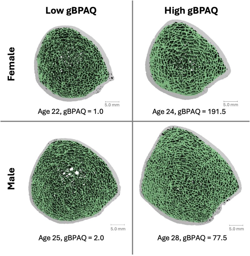
儿童期和青春期是骨骼发育的关键时期,是建立峰值骨量(PBM)的重要决定因素,是终生骨折风险的重要决定因素。本研究探讨了生长期间累积体力活动与PBM周围骨密度和微结构之间的关系。226例个体(女性142例;在桡骨远端和胫骨处使用高分辨率周边定量计算机断层扫描(HR-pQCT),在腰椎和股骨颈处使用双能x线吸收仪,对84名男性(18-35岁)周围PBM进行检查。通过骨骼特异性体力活动问卷记录生长至骨量峰值期间的体力活动。经年龄、身高和体重调整后,Spearman的部分相关性被用来确定骨骼特性和生长过程中身体活动之间的性别特异性关联。通过骨特异性体力活动问卷(gBPAQ)量化的生长期间较高的体力活动与较高的胫骨衰竭负荷和股骨颈面积骨密度相关,男女均如此(ρ = 0.27-0.38, p≤0.02)。较高的gBPAQ分数也与两性更好的骨小梁骨密度和胫骨骨体积分数相关,其中男性的相关性更强(ρ = 0.40-0.41, p
The association between physical activity during growth and bone microarchitecture at peak bone mass.
Childhood and adolescence are critical periods for skeletal development in establishing peak bone mass (PBM), an important determinant of lifelong fracture risk. This study investigates the relationship between cumulative physical activity during growth and bone density and microarchitecture attained surrounding PBM. BMD and microarchitecture properties were obtained in 226 individuals (142 females; 84 males) surrounding PBM (aged 18-35 years) using HR-pQCT at the distal radius and tibia, and DXA at the lumbar spine and femoral neck. Physical activity during growth up to PBM was captured with the bone-specific physical activity questionnaire (BPAQ). Spearman's partial correlations, adjusted for age, height, and weight were used to determine sex-specific associations between bone properties and physical activity during growth. Higher physical activity during growth quantified by the BPAQ (gBPAQ) was associated with higher tibia failure load and femoral neck areal BMD, in both sexes (ρ = 0.27-0.38, p ≤ .02). Higher gBPAQ scores were also associated with better trabecular BMD and bone volume fraction at the tibia in both sexes, where associations were stronger in males (ρ = 0.40-0.41, p < .01) than in females (ρ = 0.24-0.26, p < .05). Males additionally had significant associations with trabecular bone microarchitecture properties, including number, separation, and inhomogeneity at both the radius (ρ = 0.30-0.34, p ≤ .01) and tibia (ρ = 0.31-0.42, p ≤ .02). In contrast, gBPAQ scores were not associated with cortical bone properties at PBM for either sex or site. Physical activity during growth is associated with greater BMD and failure load at PBM, 2 predictors of lifelong fracture risk. However, compartment-specific differences indicate that trabecular bone, as opposed to cortical bone, is more responsive to physical activity during growth.
期刊介绍:
The Journal of Bone and Mineral Research (JBMR) publishes highly impactful original manuscripts, reviews, and special articles on basic, translational and clinical investigations relevant to the musculoskeletal system and mineral metabolism. Specifically, the journal is interested in original research on the biology and physiology of skeletal tissues, interdisciplinary research spanning the musculoskeletal and other systems, including but not limited to immunology, hematology, energy metabolism, cancer biology, and neurology, and systems biology topics using large scale “-omics” approaches. The journal welcomes clinical research on the pathophysiology, treatment and prevention of osteoporosis and fractures, as well as sarcopenia, disorders of bone and mineral metabolism, and rare or genetically determined bone diseases.
 求助内容:
求助内容: 应助结果提醒方式:


