Shunya Sadaki, Ryosuke Tsuji, Takuto Hayashi, Masato Watanabe, Ryoto Iwai, Gu Wenchao, Ekaterina A Semenova, Rinat I Sultanov, Andrey V Zhelankin, Edward V Generozov, Ildus I Ahmetov, Iori Sakakibara, Koichi Ojima, Hidetoshi Sakurai, Masafumi Muratani, Takashi Kudo, Satoru Takahashi, Ryo Fujita
{"title":"大型MAF转录因子重新唤醒人类骨骼肌中进化休眠的快速糖酵解IIb型肌纤维。","authors":"Shunya Sadaki, Ryosuke Tsuji, Takuto Hayashi, Masato Watanabe, Ryoto Iwai, Gu Wenchao, Ekaterina A Semenova, Rinat I Sultanov, Andrey V Zhelankin, Edward V Generozov, Ildus I Ahmetov, Iori Sakakibara, Koichi Ojima, Hidetoshi Sakurai, Masafumi Muratani, Takashi Kudo, Satoru Takahashi, Ryo Fujita","doi":"10.1186/s13395-025-00391-5","DOIUrl":null,"url":null,"abstract":"<p><strong>Background: </strong>Small mammals such as mice rely on type IIb myofibers, which express the fast-contracting myosin heavy chain isoform Myh4, to achieve rapid movements. In contrast, larger mammals, including humans, have lost MYH4 expression. Thus, they favor slower-contracting myofiber types. However, the mechanisms underlying this evolutionary shift remain unclear. We recently identified the large Maf transcription factor family (Mafa, Mafb, and Maf) as key regulators of type IIb myofiber specification in mice. In this study, we investigate whether large MAFs play a conserved role in the induction of MYH4 expression and glycolytic metabolism in human and bovine skeletal muscle.</p><p><strong>Methods: </strong>We performed adenovirus-mediated overexpression of large MAFs in iPSC-derived human myotubes and primary bovine myotubes. We subsequently quantified MYH4 expression using RT-qPCR, RNA sequencing (RNA-seq), and LC-MS/MS analysis. Glycolytic capacity was assessed using a flux analyzer and metabolic gene expression profiling. Additionally, RNA-seq analysis of human muscle biopsy samples was conducted to determine the correlations between large MAFs and the expression of MYH4 and other myosin genes, as well as their association with fast fiber composition and athletic training.</p><p><strong>Results: </strong>Overexpression of large MAFs in human and bovine myotubes robustly induced MYH4 expression, with mRNA levels increasing by 100- to 1000-fold. LC-MS/MS analysis provided clear evidence of MYH4 protein expression in human myotubes, where it was previously undetectable. RNA-seq and flux analyzer data revealed that large MAFs significantly enhanced glycolytic capacity by upregulating the expression of key genes involved in glucose metabolism. Moreover, RNA-seq analysis of human muscle biopsy samples revealed a positive correlation between MAFA, MAF, and MYH4 expression. Furthermore, MAFA and MAF expression levels were elevated in power-trained individuals, accompanied by increased expression of MYH4 and other fast myosin genes.</p><p><strong>Conclusions: </strong>Our findings establish large MAF transcription factors as key regulators of MYH4 expression and glycolytic metabolism in human skeletal muscle. This discovery provides novel insights into the evolutionary loss of type IIb myofibers in larger mammals and suggests potential strategies for enhancing muscle performance and mitigating fast-twitch fiber loss associated with aging and muscle degeneration.</p>","PeriodicalId":21747,"journal":{"name":"Skeletal Muscle","volume":"15 1","pages":"19"},"PeriodicalIF":4.4000,"publicationDate":"2025-07-26","publicationTypes":"Journal Article","fieldsOfStudy":null,"isOpenAccess":false,"openAccessPdf":"https://www.ncbi.nlm.nih.gov/pmc/articles/PMC12296675/pdf/","citationCount":"0","resultStr":"{\"title\":\"Large MAF transcription factors reawaken evolutionarily dormant fast-glycolytic type IIb myofibers in human skeletal muscle.\",\"authors\":\"Shunya Sadaki, Ryosuke Tsuji, Takuto Hayashi, Masato Watanabe, Ryoto Iwai, Gu Wenchao, Ekaterina A Semenova, Rinat I Sultanov, Andrey V Zhelankin, Edward V Generozov, Ildus I Ahmetov, Iori Sakakibara, Koichi Ojima, Hidetoshi Sakurai, Masafumi Muratani, Takashi Kudo, Satoru Takahashi, Ryo Fujita\",\"doi\":\"10.1186/s13395-025-00391-5\",\"DOIUrl\":null,\"url\":null,\"abstract\":\"<p><strong>Background: </strong>Small mammals such as mice rely on type IIb myofibers, which express the fast-contracting myosin heavy chain isoform Myh4, to achieve rapid movements. In contrast, larger mammals, including humans, have lost MYH4 expression. Thus, they favor slower-contracting myofiber types. However, the mechanisms underlying this evolutionary shift remain unclear. We recently identified the large Maf transcription factor family (Mafa, Mafb, and Maf) as key regulators of type IIb myofiber specification in mice. In this study, we investigate whether large MAFs play a conserved role in the induction of MYH4 expression and glycolytic metabolism in human and bovine skeletal muscle.</p><p><strong>Methods: </strong>We performed adenovirus-mediated overexpression of large MAFs in iPSC-derived human myotubes and primary bovine myotubes. We subsequently quantified MYH4 expression using RT-qPCR, RNA sequencing (RNA-seq), and LC-MS/MS analysis. Glycolytic capacity was assessed using a flux analyzer and metabolic gene expression profiling. Additionally, RNA-seq analysis of human muscle biopsy samples was conducted to determine the correlations between large MAFs and the expression of MYH4 and other myosin genes, as well as their association with fast fiber composition and athletic training.</p><p><strong>Results: </strong>Overexpression of large MAFs in human and bovine myotubes robustly induced MYH4 expression, with mRNA levels increasing by 100- to 1000-fold. LC-MS/MS analysis provided clear evidence of MYH4 protein expression in human myotubes, where it was previously undetectable. RNA-seq and flux analyzer data revealed that large MAFs significantly enhanced glycolytic capacity by upregulating the expression of key genes involved in glucose metabolism. Moreover, RNA-seq analysis of human muscle biopsy samples revealed a positive correlation between MAFA, MAF, and MYH4 expression. Furthermore, MAFA and MAF expression levels were elevated in power-trained individuals, accompanied by increased expression of MYH4 and other fast myosin genes.</p><p><strong>Conclusions: </strong>Our findings establish large MAF transcription factors as key regulators of MYH4 expression and glycolytic metabolism in human skeletal muscle. This discovery provides novel insights into the evolutionary loss of type IIb myofibers in larger mammals and suggests potential strategies for enhancing muscle performance and mitigating fast-twitch fiber loss associated with aging and muscle degeneration.</p>\",\"PeriodicalId\":21747,\"journal\":{\"name\":\"Skeletal Muscle\",\"volume\":\"15 1\",\"pages\":\"19\"},\"PeriodicalIF\":4.4000,\"publicationDate\":\"2025-07-26\",\"publicationTypes\":\"Journal Article\",\"fieldsOfStudy\":null,\"isOpenAccess\":false,\"openAccessPdf\":\"https://www.ncbi.nlm.nih.gov/pmc/articles/PMC12296675/pdf/\",\"citationCount\":\"0\",\"resultStr\":null,\"platform\":\"Semanticscholar\",\"paperid\":null,\"PeriodicalName\":\"Skeletal Muscle\",\"FirstCategoryId\":\"3\",\"ListUrlMain\":\"https://doi.org/10.1186/s13395-025-00391-5\",\"RegionNum\":2,\"RegionCategory\":\"医学\",\"ArticlePicture\":[],\"TitleCN\":null,\"AbstractTextCN\":null,\"PMCID\":null,\"EPubDate\":\"\",\"PubModel\":\"\",\"JCR\":\"Q2\",\"JCRName\":\"CELL BIOLOGY\",\"Score\":null,\"Total\":0}","platform":"Semanticscholar","paperid":null,"PeriodicalName":"Skeletal Muscle","FirstCategoryId":"3","ListUrlMain":"https://doi.org/10.1186/s13395-025-00391-5","RegionNum":2,"RegionCategory":"医学","ArticlePicture":[],"TitleCN":null,"AbstractTextCN":null,"PMCID":null,"EPubDate":"","PubModel":"","JCR":"Q2","JCRName":"CELL BIOLOGY","Score":null,"Total":0}
Large MAF transcription factors reawaken evolutionarily dormant fast-glycolytic type IIb myofibers in human skeletal muscle.
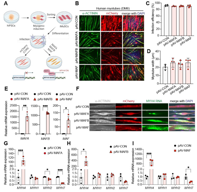
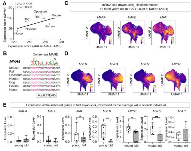
Background: Small mammals such as mice rely on type IIb myofibers, which express the fast-contracting myosin heavy chain isoform Myh4, to achieve rapid movements. In contrast, larger mammals, including humans, have lost MYH4 expression. Thus, they favor slower-contracting myofiber types. However, the mechanisms underlying this evolutionary shift remain unclear. We recently identified the large Maf transcription factor family (Mafa, Mafb, and Maf) as key regulators of type IIb myofiber specification in mice. In this study, we investigate whether large MAFs play a conserved role in the induction of MYH4 expression and glycolytic metabolism in human and bovine skeletal muscle.
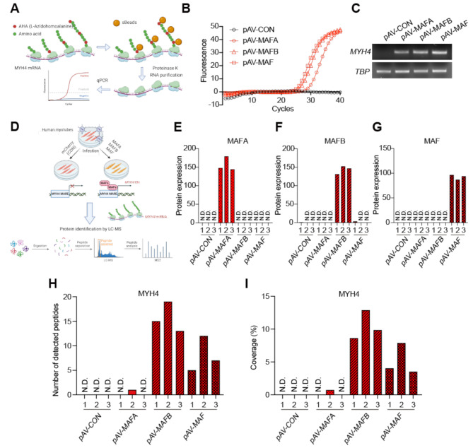
Methods: We performed adenovirus-mediated overexpression of large MAFs in iPSC-derived human myotubes and primary bovine myotubes. We subsequently quantified MYH4 expression using RT-qPCR, RNA sequencing (RNA-seq), and LC-MS/MS analysis. Glycolytic capacity was assessed using a flux analyzer and metabolic gene expression profiling. Additionally, RNA-seq analysis of human muscle biopsy samples was conducted to determine the correlations between large MAFs and the expression of MYH4 and other myosin genes, as well as their association with fast fiber composition and athletic training.
Results: Overexpression of large MAFs in human and bovine myotubes robustly induced MYH4 expression, with mRNA levels increasing by 100- to 1000-fold. LC-MS/MS analysis provided clear evidence of MYH4 protein expression in human myotubes, where it was previously undetectable. RNA-seq and flux analyzer data revealed that large MAFs significantly enhanced glycolytic capacity by upregulating the expression of key genes involved in glucose metabolism. Moreover, RNA-seq analysis of human muscle biopsy samples revealed a positive correlation between MAFA, MAF, and MYH4 expression. Furthermore, MAFA and MAF expression levels were elevated in power-trained individuals, accompanied by increased expression of MYH4 and other fast myosin genes.
Conclusions: Our findings establish large MAF transcription factors as key regulators of MYH4 expression and glycolytic metabolism in human skeletal muscle. This discovery provides novel insights into the evolutionary loss of type IIb myofibers in larger mammals and suggests potential strategies for enhancing muscle performance and mitigating fast-twitch fiber loss associated with aging and muscle degeneration.
期刊介绍:
The only open access journal in its field, Skeletal Muscle publishes novel, cutting-edge research and technological advancements that investigate the molecular mechanisms underlying the biology of skeletal muscle. Reflecting the breadth of research in this area, the journal welcomes manuscripts about the development, metabolism, the regulation of mass and function, aging, degeneration, dystrophy and regeneration of skeletal muscle, with an emphasis on understanding adult skeletal muscle, its maintenance, and its interactions with non-muscle cell types and regulatory modulators.
Main areas of interest include:
-differentiation of skeletal muscle-
atrophy and hypertrophy of skeletal muscle-
aging of skeletal muscle-
regeneration and degeneration of skeletal muscle-
biology of satellite and satellite-like cells-
dystrophic degeneration of skeletal muscle-
energy and glucose homeostasis in skeletal muscle-
non-dystrophic genetic diseases of skeletal muscle, such as Spinal Muscular Atrophy and myopathies-
maintenance of neuromuscular junctions-
roles of ryanodine receptors and calcium signaling in skeletal muscle-
roles of nuclear receptors in skeletal muscle-
roles of GPCRs and GPCR signaling in skeletal muscle-
other relevant aspects of skeletal muscle biology.
In addition, articles on translational clinical studies that address molecular and cellular mechanisms of skeletal muscle will be published. Case reports are also encouraged for submission.
Skeletal Muscle reflects the breadth of research on skeletal muscle and bridges gaps between diverse areas of science for example cardiac cell biology and neurobiology, which share common features with respect to cell differentiation, excitatory membranes, cell-cell communication, and maintenance. Suitable articles are model and mechanism-driven, and apply statistical principles where appropriate; purely descriptive studies are of lesser interest.
 求助内容:
求助内容: 应助结果提醒方式:


