Pedro Christian Aravena, Mario E Flores, Larissa Córdova Turones, Francisca Pavicic, Pamela Ehrenfeld
{"title":"用聚己内酯三维根复制品保存牙槽嵴:一个病例报告的放射学和组织学评价。","authors":"Pedro Christian Aravena, Mario E Flores, Larissa Córdova Turones, Francisca Pavicic, Pamela Ehrenfeld","doi":"10.3390/reports8020092","DOIUrl":null,"url":null,"abstract":"<p><p><b>Background and Clinical Significance:</b> To describe the effectiveness of alveolar ridge preservation under the radiological and histological analysis of a customized resorbable scaffold three-dimensionally printed with polycaprolactone (PCL) reinforced with a coating of a copolymer of polycaprolactone-block-polyethylene glycol (PCL-PEG) by electrospray. <b>Case Presentation:</b> A 62-year-old male with vertical root fractures of teeth #14 and #15. From the cone beam CT (CBCT) image, the scaffold root replicas were designed with the shape of the roots and printed with PCL coated with PCL-PEG by electrospray. The scaffold was inserted into the alveolar bone and maintained with a tension-free flap closure. After six months, a CBCT of the surgical site and histological analysis of a bone sample at the dental implant installation site were performed. After 6 months, the wound in tooth #14 was closed, clinically proving no adverse reaction or complications. The histological analysis of the bone sample showed new bone formation with lamellar structure, Haversian canal structure, and osteocyte spaces. However, the scaffold in tooth #15 was exposed and not osseointegrated, and it was covered with membranous tissue. Histologically, the sample showed tissue compatible with lax connective tissue with mixed inflammatory infiltrate. In tooth #14, the dental implant presented an insertion torque >35 Ncm and was rehabilitated three months after its installation. <b>Conclusions:</b> Three-dimensional printed PCL scaffolds showed the ability to regenerate vital and functional bone with osseointegration capability for maxillary bone regeneration and oral rehabilitation based on dental implants. A case of inadequate scaffold osseointegration accompanied by lax connective tissue formation is shown.</p>","PeriodicalId":74664,"journal":{"name":"Reports (MDPI)","volume":"8 2","pages":""},"PeriodicalIF":0.8000,"publicationDate":"2025-06-09","publicationTypes":"Journal Article","fieldsOfStudy":null,"isOpenAccess":false,"openAccessPdf":"https://www.ncbi.nlm.nih.gov/pmc/articles/PMC12196692/pdf/","citationCount":"0","resultStr":"{\"title\":\"Alveolar Ridge Preservation Using Three-Dimensional Root Replicas of Polycaprolactone: A Radiological and Histological Evaluation of a Case Report.\",\"authors\":\"Pedro Christian Aravena, Mario E Flores, Larissa Córdova Turones, Francisca Pavicic, Pamela Ehrenfeld\",\"doi\":\"10.3390/reports8020092\",\"DOIUrl\":null,\"url\":null,\"abstract\":\"<p><p><b>Background and Clinical Significance:</b> To describe the effectiveness of alveolar ridge preservation under the radiological and histological analysis of a customized resorbable scaffold three-dimensionally printed with polycaprolactone (PCL) reinforced with a coating of a copolymer of polycaprolactone-block-polyethylene glycol (PCL-PEG) by electrospray. <b>Case Presentation:</b> A 62-year-old male with vertical root fractures of teeth #14 and #15. From the cone beam CT (CBCT) image, the scaffold root replicas were designed with the shape of the roots and printed with PCL coated with PCL-PEG by electrospray. The scaffold was inserted into the alveolar bone and maintained with a tension-free flap closure. After six months, a CBCT of the surgical site and histological analysis of a bone sample at the dental implant installation site were performed. After 6 months, the wound in tooth #14 was closed, clinically proving no adverse reaction or complications. The histological analysis of the bone sample showed new bone formation with lamellar structure, Haversian canal structure, and osteocyte spaces. However, the scaffold in tooth #15 was exposed and not osseointegrated, and it was covered with membranous tissue. Histologically, the sample showed tissue compatible with lax connective tissue with mixed inflammatory infiltrate. In tooth #14, the dental implant presented an insertion torque >35 Ncm and was rehabilitated three months after its installation. <b>Conclusions:</b> Three-dimensional printed PCL scaffolds showed the ability to regenerate vital and functional bone with osseointegration capability for maxillary bone regeneration and oral rehabilitation based on dental implants. A case of inadequate scaffold osseointegration accompanied by lax connective tissue formation is shown.</p>\",\"PeriodicalId\":74664,\"journal\":{\"name\":\"Reports (MDPI)\",\"volume\":\"8 2\",\"pages\":\"\"},\"PeriodicalIF\":0.8000,\"publicationDate\":\"2025-06-09\",\"publicationTypes\":\"Journal Article\",\"fieldsOfStudy\":null,\"isOpenAccess\":false,\"openAccessPdf\":\"https://www.ncbi.nlm.nih.gov/pmc/articles/PMC12196692/pdf/\",\"citationCount\":\"0\",\"resultStr\":null,\"platform\":\"Semanticscholar\",\"paperid\":null,\"PeriodicalName\":\"Reports (MDPI)\",\"FirstCategoryId\":\"1085\",\"ListUrlMain\":\"https://doi.org/10.3390/reports8020092\",\"RegionNum\":0,\"RegionCategory\":null,\"ArticlePicture\":[],\"TitleCN\":null,\"AbstractTextCN\":null,\"PMCID\":null,\"EPubDate\":\"\",\"PubModel\":\"\",\"JCR\":\"Q3\",\"JCRName\":\"MEDICINE, GENERAL & INTERNAL\",\"Score\":null,\"Total\":0}","platform":"Semanticscholar","paperid":null,"PeriodicalName":"Reports (MDPI)","FirstCategoryId":"1085","ListUrlMain":"https://doi.org/10.3390/reports8020092","RegionNum":0,"RegionCategory":null,"ArticlePicture":[],"TitleCN":null,"AbstractTextCN":null,"PMCID":null,"EPubDate":"","PubModel":"","JCR":"Q3","JCRName":"MEDICINE, GENERAL & INTERNAL","Score":null,"Total":0}
Alveolar Ridge Preservation Using Three-Dimensional Root Replicas of Polycaprolactone: A Radiological and Histological Evaluation of a Case Report.
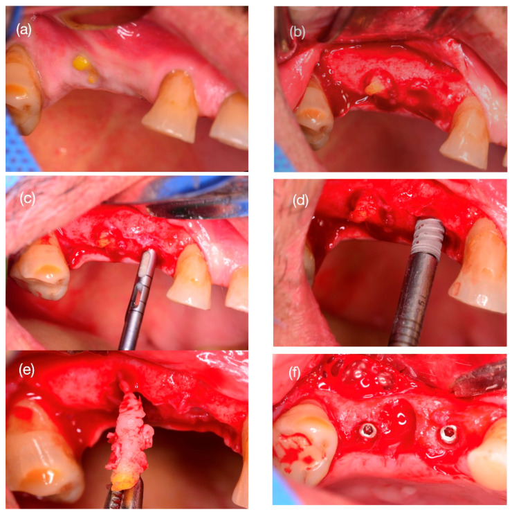
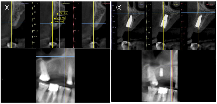
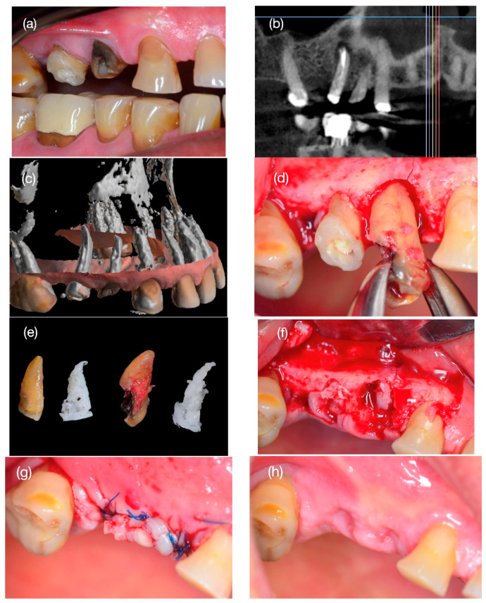
Background and Clinical Significance: To describe the effectiveness of alveolar ridge preservation under the radiological and histological analysis of a customized resorbable scaffold three-dimensionally printed with polycaprolactone (PCL) reinforced with a coating of a copolymer of polycaprolactone-block-polyethylene glycol (PCL-PEG) by electrospray. Case Presentation: A 62-year-old male with vertical root fractures of teeth #14 and #15. From the cone beam CT (CBCT) image, the scaffold root replicas were designed with the shape of the roots and printed with PCL coated with PCL-PEG by electrospray. The scaffold was inserted into the alveolar bone and maintained with a tension-free flap closure. After six months, a CBCT of the surgical site and histological analysis of a bone sample at the dental implant installation site were performed. After 6 months, the wound in tooth #14 was closed, clinically proving no adverse reaction or complications. The histological analysis of the bone sample showed new bone formation with lamellar structure, Haversian canal structure, and osteocyte spaces. However, the scaffold in tooth #15 was exposed and not osseointegrated, and it was covered with membranous tissue. Histologically, the sample showed tissue compatible with lax connective tissue with mixed inflammatory infiltrate. In tooth #14, the dental implant presented an insertion torque >35 Ncm and was rehabilitated three months after its installation. Conclusions: Three-dimensional printed PCL scaffolds showed the ability to regenerate vital and functional bone with osseointegration capability for maxillary bone regeneration and oral rehabilitation based on dental implants. A case of inadequate scaffold osseointegration accompanied by lax connective tissue formation is shown.

 求助内容:
求助内容: 应助结果提醒方式:


