{"title":"细胞病理学家在放射科医生的协助下对甲状腺外头颈部病变进行细针穿刺细胞学诊断的准确性:一项单中心研究。","authors":"Busra Yaprak Bayrak, Nadir Paksoy","doi":"10.25259/Cytojournal_247_2024","DOIUrl":null,"url":null,"abstract":"<p><strong>Objective: </strong>In recent years, several publications have described the use of ultrasound-guided fine-needle aspiration (FNA) by cytopathologists to achieve better diagnostic accuracy. Some cytopathologists enroll in courses to learn and apply ultrasound (US) guidance themselves. However, no standard procedure has been established that cytopathologists can follow to perform US for FNA. Alternatively, FNA can be a useful tool when cytopathologists collaborate with radiologists. Here, we aimed to evaluate the diagnostic accuracy of FNA for non-thyroidal head-and-neck masses retrieved by a cytopathologist with US guidance provided by a radiologist.</p><p><strong>Material and methods: </strong>The FNA results for non-thyroidal head-and-neck masses at a private clinic using the Scandinavian FNA model with radiologist‒cytopathologist collaboration were compared with the histopathology results.</p><p><strong>Results: </strong>In all, 1890 patients who underwent FNA were identified, among whom 1435 (76%) also had histopathological results. Non-cystic lesions were obtained from lymph nodes (LNs), salivary glands, and soft tissue, while the other lesions were cystic in nature. For FNA, the accuracy was 99.4%, the sensitivity was 99.6%, the specificity was 99.3%, the positive predictive value was 99.3%, and the negative predictive value was 99.6%. No FNA results were non-diagnostic. Surgical follow-up revealed that only eight of the 1435 assessments (0.5%), all performed for LN lesions, yielded false-negative or false-positive results.</p><p><strong>Conclusion: </strong>The present study is based on single-center observations. The use of FNA, when performed by a specialized cytopathologist and with US assistance from a radiologist, produces accurate results and sufficient material for analysis, especially for LNs in extrathyroidal head-and-neck lesions. This study also reveals that the technique is a low-cost and effective process. The way in which FNA is presented here indicates that this procedure would be useful and ideal for any health service.</p>","PeriodicalId":49082,"journal":{"name":"Cytojournal","volume":"22 ","pages":"57"},"PeriodicalIF":3.1000,"publicationDate":"2025-06-02","publicationTypes":"Journal Article","fieldsOfStudy":null,"isOpenAccess":false,"openAccessPdf":"https://www.ncbi.nlm.nih.gov/pmc/articles/PMC12289109/pdf/","citationCount":"0","resultStr":"{\"title\":\"Diagnostic accuracy of fine-needle aspiration cytology for extrathyroidal head-and-neck lesions performed by a cytopathologist with the assistance of radiologist: A single-center study.\",\"authors\":\"Busra Yaprak Bayrak, Nadir Paksoy\",\"doi\":\"10.25259/Cytojournal_247_2024\",\"DOIUrl\":null,\"url\":null,\"abstract\":\"<p><strong>Objective: </strong>In recent years, several publications have described the use of ultrasound-guided fine-needle aspiration (FNA) by cytopathologists to achieve better diagnostic accuracy. Some cytopathologists enroll in courses to learn and apply ultrasound (US) guidance themselves. However, no standard procedure has been established that cytopathologists can follow to perform US for FNA. Alternatively, FNA can be a useful tool when cytopathologists collaborate with radiologists. Here, we aimed to evaluate the diagnostic accuracy of FNA for non-thyroidal head-and-neck masses retrieved by a cytopathologist with US guidance provided by a radiologist.</p><p><strong>Material and methods: </strong>The FNA results for non-thyroidal head-and-neck masses at a private clinic using the Scandinavian FNA model with radiologist‒cytopathologist collaboration were compared with the histopathology results.</p><p><strong>Results: </strong>In all, 1890 patients who underwent FNA were identified, among whom 1435 (76%) also had histopathological results. Non-cystic lesions were obtained from lymph nodes (LNs), salivary glands, and soft tissue, while the other lesions were cystic in nature. For FNA, the accuracy was 99.4%, the sensitivity was 99.6%, the specificity was 99.3%, the positive predictive value was 99.3%, and the negative predictive value was 99.6%. No FNA results were non-diagnostic. Surgical follow-up revealed that only eight of the 1435 assessments (0.5%), all performed for LN lesions, yielded false-negative or false-positive results.</p><p><strong>Conclusion: </strong>The present study is based on single-center observations. The use of FNA, when performed by a specialized cytopathologist and with US assistance from a radiologist, produces accurate results and sufficient material for analysis, especially for LNs in extrathyroidal head-and-neck lesions. This study also reveals that the technique is a low-cost and effective process. The way in which FNA is presented here indicates that this procedure would be useful and ideal for any health service.</p>\",\"PeriodicalId\":49082,\"journal\":{\"name\":\"Cytojournal\",\"volume\":\"22 \",\"pages\":\"57\"},\"PeriodicalIF\":3.1000,\"publicationDate\":\"2025-06-02\",\"publicationTypes\":\"Journal Article\",\"fieldsOfStudy\":null,\"isOpenAccess\":false,\"openAccessPdf\":\"https://www.ncbi.nlm.nih.gov/pmc/articles/PMC12289109/pdf/\",\"citationCount\":\"0\",\"resultStr\":null,\"platform\":\"Semanticscholar\",\"paperid\":null,\"PeriodicalName\":\"Cytojournal\",\"FirstCategoryId\":\"3\",\"ListUrlMain\":\"https://doi.org/10.25259/Cytojournal_247_2024\",\"RegionNum\":4,\"RegionCategory\":\"医学\",\"ArticlePicture\":[],\"TitleCN\":null,\"AbstractTextCN\":null,\"PMCID\":null,\"EPubDate\":\"2025/1/1 0:00:00\",\"PubModel\":\"eCollection\",\"JCR\":\"Q2\",\"JCRName\":\"PATHOLOGY\",\"Score\":null,\"Total\":0}","platform":"Semanticscholar","paperid":null,"PeriodicalName":"Cytojournal","FirstCategoryId":"3","ListUrlMain":"https://doi.org/10.25259/Cytojournal_247_2024","RegionNum":4,"RegionCategory":"医学","ArticlePicture":[],"TitleCN":null,"AbstractTextCN":null,"PMCID":null,"EPubDate":"2025/1/1 0:00:00","PubModel":"eCollection","JCR":"Q2","JCRName":"PATHOLOGY","Score":null,"Total":0}
Diagnostic accuracy of fine-needle aspiration cytology for extrathyroidal head-and-neck lesions performed by a cytopathologist with the assistance of radiologist: A single-center study.
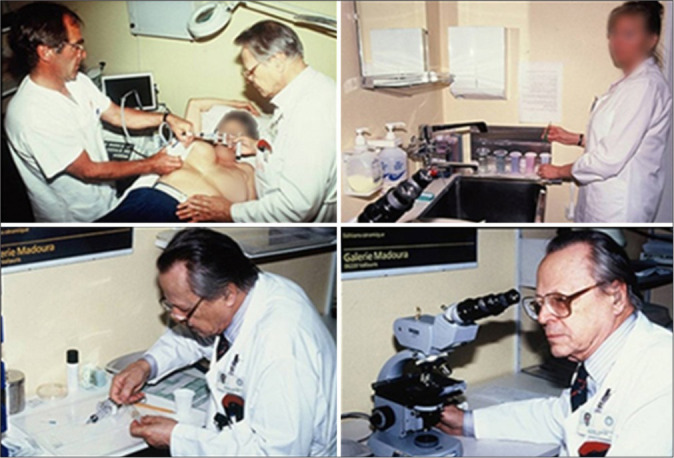
Objective: In recent years, several publications have described the use of ultrasound-guided fine-needle aspiration (FNA) by cytopathologists to achieve better diagnostic accuracy. Some cytopathologists enroll in courses to learn and apply ultrasound (US) guidance themselves. However, no standard procedure has been established that cytopathologists can follow to perform US for FNA. Alternatively, FNA can be a useful tool when cytopathologists collaborate with radiologists. Here, we aimed to evaluate the diagnostic accuracy of FNA for non-thyroidal head-and-neck masses retrieved by a cytopathologist with US guidance provided by a radiologist.
Material and methods: The FNA results for non-thyroidal head-and-neck masses at a private clinic using the Scandinavian FNA model with radiologist‒cytopathologist collaboration were compared with the histopathology results.
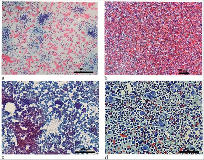
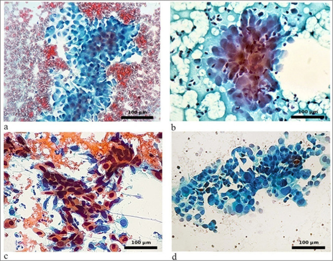
Results: In all, 1890 patients who underwent FNA were identified, among whom 1435 (76%) also had histopathological results. Non-cystic lesions were obtained from lymph nodes (LNs), salivary glands, and soft tissue, while the other lesions were cystic in nature. For FNA, the accuracy was 99.4%, the sensitivity was 99.6%, the specificity was 99.3%, the positive predictive value was 99.3%, and the negative predictive value was 99.6%. No FNA results were non-diagnostic. Surgical follow-up revealed that only eight of the 1435 assessments (0.5%), all performed for LN lesions, yielded false-negative or false-positive results.
Conclusion: The present study is based on single-center observations. The use of FNA, when performed by a specialized cytopathologist and with US assistance from a radiologist, produces accurate results and sufficient material for analysis, especially for LNs in extrathyroidal head-and-neck lesions. This study also reveals that the technique is a low-cost and effective process. The way in which FNA is presented here indicates that this procedure would be useful and ideal for any health service.
期刊介绍:
The CytoJournal is an open-access peer-reviewed journal committed to publishing high-quality articles in the field of Diagnostic Cytopathology including Molecular aspects. The journal is owned by the Cytopathology Foundation and published by the Scientific Scholar.
 求助内容:
求助内容: 应助结果提醒方式:


