{"title":"固定正畸治疗后牙髓结石的形成:拔牙和非拔牙方法的全景x线摄影比较","authors":"Kosar Gholinezhad, Hakimeh Ghorbani, Seyedali Seyedmajidi, Sedigheh Sheikhzadeh, Manouchehr Rahmati Kamel","doi":"10.1002/cre2.70181","DOIUrl":null,"url":null,"abstract":"<div>\n \n \n <section>\n \n <h3> Objective</h3>\n \n <p>The impact of orthodontic forces on pulp stone formation has been the focus of several studies. Given that orthodontic extractions typically involve the application of greater forces to the teeth, the aim of this study was to compare the extent of pulp stone formation in the molar teeth of patients undergoing orthodontic treatment with and without extractions.</p>\n </section>\n \n <section>\n \n <h3> Material and Methods</h3>\n \n <p>In this retrospective observational study, panoramic radiographs of 80 orthodontic patients taken between 2014 and 2020 (equally divided into extraction and non-extraction groups) who had a full set of permanent molars were analyzed before and after orthodontic treatment to assess the formation of pulp stones in the pulp chambers of the molar teeth (640 molars). Data were analyzed using the Chi-square and McNemar tests with a significance level set at <i>p</i> < 0.05, using SPSS software.</p>\n </section>\n \n <section>\n \n <h3> Results</h3>\n \n <p>The frequency of pulp stone formation significantly increased in both the extraction and non-extraction groups following fixed orthodontic treatment (<i>p</i> < 0.001 and <i>p</i> = 0.02, respectively). However, no statistically significant difference was observed in the extent of pulp stone formation between the two groups (<i>p</i> = 0.09). The frequency of patients exhibiting pulp stone formation did not differ significantly by gender in either the extraction or non-extraction treatment groups (<i>p</i> = 0.392 and <i>p</i> = 0.451, respectively). In the extraction group, the prevalence of pulp stones was significantly higher in the first molar compared to the second molar (<i>p</i> = 0.001). In contrast, no significant difference was found between the first and second molars in the non-extraction group (<i>p</i> = 0.108). Additionally, no correlation was found between the frequency of pulp stone formation and jaw type (maxilla or mandible) in either group (<i>p</i> > 0.05).</p>\n </section>\n \n <section>\n \n <h3> Conclusions</h3>\n \n <p>Fixed orthodontic treatment is associated with increased pulp stone formation, regardless of whether extractions are performed. These findings may help clinicians in the early identification and monitoring of at-risk teeth.</p>\n </section>\n </div>","PeriodicalId":10203,"journal":{"name":"Clinical and Experimental Dental Research","volume":"11 4","pages":""},"PeriodicalIF":2.2000,"publicationDate":"2025-07-24","publicationTypes":"Journal Article","fieldsOfStudy":null,"isOpenAccess":false,"openAccessPdf":"https://onlinelibrary.wiley.com/doi/epdf/10.1002/cre2.70181","citationCount":"0","resultStr":"{\"title\":\"Pulp Stone Formation Following Fixed Orthodontic Treatment: A Panoramic Radiographic Comparison of Extraction and Non-Extraction Approaches\",\"authors\":\"Kosar Gholinezhad, Hakimeh Ghorbani, Seyedali Seyedmajidi, Sedigheh Sheikhzadeh, Manouchehr Rahmati Kamel\",\"doi\":\"10.1002/cre2.70181\",\"DOIUrl\":null,\"url\":null,\"abstract\":\"<div>\\n \\n \\n <section>\\n \\n <h3> Objective</h3>\\n \\n <p>The impact of orthodontic forces on pulp stone formation has been the focus of several studies. Given that orthodontic extractions typically involve the application of greater forces to the teeth, the aim of this study was to compare the extent of pulp stone formation in the molar teeth of patients undergoing orthodontic treatment with and without extractions.</p>\\n </section>\\n \\n <section>\\n \\n <h3> Material and Methods</h3>\\n \\n <p>In this retrospective observational study, panoramic radiographs of 80 orthodontic patients taken between 2014 and 2020 (equally divided into extraction and non-extraction groups) who had a full set of permanent molars were analyzed before and after orthodontic treatment to assess the formation of pulp stones in the pulp chambers of the molar teeth (640 molars). Data were analyzed using the Chi-square and McNemar tests with a significance level set at <i>p</i> < 0.05, using SPSS software.</p>\\n </section>\\n \\n <section>\\n \\n <h3> Results</h3>\\n \\n <p>The frequency of pulp stone formation significantly increased in both the extraction and non-extraction groups following fixed orthodontic treatment (<i>p</i> < 0.001 and <i>p</i> = 0.02, respectively). However, no statistically significant difference was observed in the extent of pulp stone formation between the two groups (<i>p</i> = 0.09). The frequency of patients exhibiting pulp stone formation did not differ significantly by gender in either the extraction or non-extraction treatment groups (<i>p</i> = 0.392 and <i>p</i> = 0.451, respectively). In the extraction group, the prevalence of pulp stones was significantly higher in the first molar compared to the second molar (<i>p</i> = 0.001). In contrast, no significant difference was found between the first and second molars in the non-extraction group (<i>p</i> = 0.108). Additionally, no correlation was found between the frequency of pulp stone formation and jaw type (maxilla or mandible) in either group (<i>p</i> > 0.05).</p>\\n </section>\\n \\n <section>\\n \\n <h3> Conclusions</h3>\\n \\n <p>Fixed orthodontic treatment is associated with increased pulp stone formation, regardless of whether extractions are performed. These findings may help clinicians in the early identification and monitoring of at-risk teeth.</p>\\n </section>\\n </div>\",\"PeriodicalId\":10203,\"journal\":{\"name\":\"Clinical and Experimental Dental Research\",\"volume\":\"11 4\",\"pages\":\"\"},\"PeriodicalIF\":2.2000,\"publicationDate\":\"2025-07-24\",\"publicationTypes\":\"Journal Article\",\"fieldsOfStudy\":null,\"isOpenAccess\":false,\"openAccessPdf\":\"https://onlinelibrary.wiley.com/doi/epdf/10.1002/cre2.70181\",\"citationCount\":\"0\",\"resultStr\":null,\"platform\":\"Semanticscholar\",\"paperid\":null,\"PeriodicalName\":\"Clinical and Experimental Dental Research\",\"FirstCategoryId\":\"1085\",\"ListUrlMain\":\"https://onlinelibrary.wiley.com/doi/10.1002/cre2.70181\",\"RegionNum\":0,\"RegionCategory\":null,\"ArticlePicture\":[],\"TitleCN\":null,\"AbstractTextCN\":null,\"PMCID\":null,\"EPubDate\":\"\",\"PubModel\":\"\",\"JCR\":\"Q3\",\"JCRName\":\"DENTISTRY, ORAL SURGERY & MEDICINE\",\"Score\":null,\"Total\":0}","platform":"Semanticscholar","paperid":null,"PeriodicalName":"Clinical and Experimental Dental Research","FirstCategoryId":"1085","ListUrlMain":"https://onlinelibrary.wiley.com/doi/10.1002/cre2.70181","RegionNum":0,"RegionCategory":null,"ArticlePicture":[],"TitleCN":null,"AbstractTextCN":null,"PMCID":null,"EPubDate":"","PubModel":"","JCR":"Q3","JCRName":"DENTISTRY, ORAL SURGERY & MEDICINE","Score":null,"Total":0}
Pulp Stone Formation Following Fixed Orthodontic Treatment: A Panoramic Radiographic Comparison of Extraction and Non-Extraction Approaches
Objective
The impact of orthodontic forces on pulp stone formation has been the focus of several studies. Given that orthodontic extractions typically involve the application of greater forces to the teeth, the aim of this study was to compare the extent of pulp stone formation in the molar teeth of patients undergoing orthodontic treatment with and without extractions.
Material and Methods
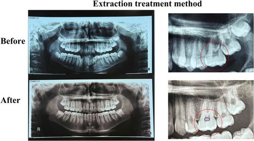
In this retrospective observational study, panoramic radiographs of 80 orthodontic patients taken between 2014 and 2020 (equally divided into extraction and non-extraction groups) who had a full set of permanent molars were analyzed before and after orthodontic treatment to assess the formation of pulp stones in the pulp chambers of the molar teeth (640 molars). Data were analyzed using the Chi-square and McNemar tests with a significance level set at p < 0.05, using SPSS software.
Results
The frequency of pulp stone formation significantly increased in both the extraction and non-extraction groups following fixed orthodontic treatment (p < 0.001 and p = 0.02, respectively). However, no statistically significant difference was observed in the extent of pulp stone formation between the two groups (p = 0.09). The frequency of patients exhibiting pulp stone formation did not differ significantly by gender in either the extraction or non-extraction treatment groups (p = 0.392 and p = 0.451, respectively). In the extraction group, the prevalence of pulp stones was significantly higher in the first molar compared to the second molar (p = 0.001). In contrast, no significant difference was found between the first and second molars in the non-extraction group (p = 0.108). Additionally, no correlation was found between the frequency of pulp stone formation and jaw type (maxilla or mandible) in either group (p > 0.05).
Conclusions
Fixed orthodontic treatment is associated with increased pulp stone formation, regardless of whether extractions are performed. These findings may help clinicians in the early identification and monitoring of at-risk teeth.
期刊介绍:
Clinical and Experimental Dental Research aims to provide open access peer-reviewed publications of high scientific quality representing original clinical, diagnostic or experimental work within all disciplines and fields of oral medicine and dentistry. The scope of Clinical and Experimental Dental Research comprises original research material on the anatomy, physiology and pathology of oro-facial, oro-pharyngeal and maxillofacial tissues, and functions and dysfunctions within the stomatognathic system, and the epidemiology, aetiology, prevention, diagnosis, prognosis and therapy of diseases and conditions that have an effect on the homeostasis of the mouth, jaws, and closely associated structures, as well as the healing and regeneration and the clinical aspects of replacement of hard and soft tissues with biomaterials, and the rehabilitation of stomatognathic functions. Studies that bring new knowledge on how to advance health on the individual or public health levels, including interactions between oral and general health and ill-health are welcome.

 求助内容:
求助内容: 应助结果提醒方式:


