MRI作为多发性硬化症间充质干细胞治疗的关键评估指标:一项系统综述和荟萃分析
IF 3.3
3区 医学
Q2 CLINICAL NEUROLOGY
引用次数: 0
摘要
多发性硬化症(MS)是一种慢性、免疫介导的疾病,以炎症、脱髓鞘和神经变性为特征,需要再生治疗。间充质干细胞(MSC)治疗具有免疫调节和神经保护的潜力,但临床评价具有挑战性。该系统综述和荟萃分析在PROSPERO (CRD420251017175)注册,遵循PRISMA 2020指南,评估MRI在评估ms的MSC治疗中的作用。我们检索了PubMed, Embase, Scopus, Web of Science和Cochrane Library,筛选了1687条记录。纳入9项同行评议的临床研究(n = 7-48例MS患者)。使用Review Manager 5.1对MRI模式(如T1/ t2加权、弥散张量成像)和结果(如病变负荷、髓鞘再生)进行叙述性和定量分析。结果常规MRI检测到病变负荷和炎症的短期减轻,而先进技术显示微结构修复,特别是鞘内MSCs。四项研究的探索性荟萃分析发现T2病变体积显著减少(平均差值-5.12 mm³,95% CI -9.65至-0.59,P = 0.03, I²= 93%),无新T2病变的可能性更高(风险比1.69,95% CI 1.31-2.19, P <;0.0001, i²=0 %)。高异质性和小样本量限制了研究结果。结论mri可捕捉病变动态,有望作为MSC治疗MS疗效的生物标志物。需要更大规模的标准化试验来解决方法上的不一致并验证研究结果。这项研究独特地强调了mri的作用,包括先进的模式,作为MS中MSC治疗的主要结果测量,突出了研究中成像标准化的差距本文章由计算机程序翻译,如有差异,请以英文原文为准。
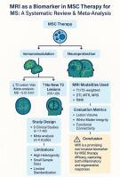
The Role of MRI as a key evaluator of mesenchymal stem Cell Therapy in Multiple Sclerosis: A systematic review and meta-analysis
Background
Multiple Sclerosis (MS) is a chronic, immune-mediated disorder marked by inflammation, demyelination, and neurodegeneration, necessitating regenerative therapies. Mesenchymal stem cell (MSC) therapy offers immunomodulatory and neuroprotective potential, but clinical evaluation is challenging.
Methods
This systematic review and meta-analysis, registered on PROSPERO (CRD420251017175) and following PRISMA 2020 guidelines, evaluated MRI’s role in assessing MSC therapy for MS. We searched PubMed, Embase, Scopus, Web of Science, and Cochrane Library, screening 1687 records. Nine peer-reviewed clinical studies (n = 7–48 MS patients) were included. MRI modalities (e.g., T1/T2-weighted, diffusion tensor imaging) and outcomes (e.g., lesion load, remyelination) were analyzed narratively and quantitatively using Review Manager 5.1.
Results
Conventional MRI detected short-term reductions in lesion load and inflammation, while advanced techniques showed microstructural repair, notably with intrathecal MSCs. An exploratory Meta-analysis of four studies found a significant T2 lesion volume decrease (mean difference -5.12 mm³, 95 % CI -9.65 to -0.59, P = 0.03, I²=93 %) and higher likelihood of no new T2 lesions (risk ratio 1.69, 95 % CI 1.31–2.19, P < 0.0001, I²=0 %). High heterogeneity and small sample sizes limited findings.
Conclusion
MRI shows promise as a biomarker for MSC therapy efficacy in MS, capturing lesion dynamics. Larger, standardized trials are needed to address methodological inconsistencies and validate findings. This study uniquely emphasizes the role of MRI—including advanced modalities—as a primary outcome measure for MSC therapy in MS, highlighting gaps in imaging standardization across studies
求助全文
通过发布文献求助,成功后即可免费获取论文全文。
去求助
来源期刊
Journal of Neuroradiology
医学-核医学
CiteScore
6.10
自引率
5.70%
发文量
142
审稿时长
6-12 weeks
期刊介绍:
The Journal of Neuroradiology is a peer-reviewed journal, publishing worldwide clinical and basic research in the field of diagnostic and Interventional neuroradiology, translational and molecular neuroimaging, and artificial intelligence in neuroradiology.
The Journal of Neuroradiology considers for publication articles, reviews, technical notes and letters to the editors (correspondence section), provided that the methodology and scientific content are of high quality, and that the results will have substantial clinical impact and/or physiological importance.
 求助内容:
求助内容: 应助结果提醒方式:


