Youngsub Eom, Eunheh Koh, Hye-Si Park, Hae-Chul Park, Jong Suk Song, Jeffrey H. Boatright, John M. Nickerson, Suhyun Kim
{"title":"红锥体消融斑马鱼幼体的一种新型色觉视运动反应试验评价","authors":"Youngsub Eom, Eunheh Koh, Hye-Si Park, Hae-Chul Park, Jong Suk Song, Jeffrey H. Boatright, John M. Nickerson, Suhyun Kim","doi":"10.1038/s41684-025-01586-5","DOIUrl":null,"url":null,"abstract":"<p>Zebrafish are widely used to investigate visual function, owing to their well-defined retinal organization and optical transparency during early developmental stages. Here, we evaluate the effectiveness of a color vision optomotor response (CV-OMR) assay for assessing zebrafish with red cone ablation. Tg (thrb:gal4;UAS:epNTR-p2a-mCherry) transgenic zebrafish were used to ablate red cones via the addition of metronidazole (MTZ). Transgenic zebrafish larvae were treated with MTZ for 0, 12 and 24 h at 5 days post-fertilization, resulting in Tg(+)MTZ(−), Tg(+)MTZ(+)12 h and Tg(+)MTZ(+)24 h groups, respectively. The areas of mCherry-expressing cells, representing red cones in tissue sections, were compared. The mean mCherry expression area was smallest in the Tg(+)MTZ(+)24 h group (16.5 ± 7.6 μm<sup>2</sup>), followed by the Tg(+)MTZ(+)12 h (404.1 ± 200.9 μm<sup>2</sup>) and Tg(+)MTZ(−) groups (1,066.6 ± 252.2 μm<sup>2</sup>; <i>P</i> < 0.001). At 6 days post-fertilization, zebrafish larvae were evaluated using the CV-OMR assay comprising two colors. Results were reported as the area under the curve of the ratio of larvae at the starting point curve. The ratio of larvae at the starting point decreased most rapidly in the Tg(+)MTZ(−) group and most slowly in the Tg(+)MTZ(+)24 h group. There were significant differences in the area under the curve of the ratio of larvae at the starting point curve among the three groups. In conclusion, the CV-OMR assay demonstrated the ability to differentiate color vision impairment based on the extent of red cone ablation. The CV-OMR assay used in this study may prove valuable for evaluating color vision in zebrafish with various eye diseases.</p>","PeriodicalId":17936,"journal":{"name":"Lab Animal","volume":"214 1","pages":""},"PeriodicalIF":3.9000,"publicationDate":"2025-07-25","publicationTypes":"Journal Article","fieldsOfStudy":null,"isOpenAccess":false,"openAccessPdf":"","citationCount":"0","resultStr":"{\"title\":\"Assessment of a novel color vision optomotor response assay in zebrafish larvae with red cone ablation\",\"authors\":\"Youngsub Eom, Eunheh Koh, Hye-Si Park, Hae-Chul Park, Jong Suk Song, Jeffrey H. Boatright, John M. Nickerson, Suhyun Kim\",\"doi\":\"10.1038/s41684-025-01586-5\",\"DOIUrl\":null,\"url\":null,\"abstract\":\"<p>Zebrafish are widely used to investigate visual function, owing to their well-defined retinal organization and optical transparency during early developmental stages. Here, we evaluate the effectiveness of a color vision optomotor response (CV-OMR) assay for assessing zebrafish with red cone ablation. Tg (thrb:gal4;UAS:epNTR-p2a-mCherry) transgenic zebrafish were used to ablate red cones via the addition of metronidazole (MTZ). Transgenic zebrafish larvae were treated with MTZ for 0, 12 and 24 h at 5 days post-fertilization, resulting in Tg(+)MTZ(−), Tg(+)MTZ(+)12 h and Tg(+)MTZ(+)24 h groups, respectively. The areas of mCherry-expressing cells, representing red cones in tissue sections, were compared. The mean mCherry expression area was smallest in the Tg(+)MTZ(+)24 h group (16.5 ± 7.6 μm<sup>2</sup>), followed by the Tg(+)MTZ(+)12 h (404.1 ± 200.9 μm<sup>2</sup>) and Tg(+)MTZ(−) groups (1,066.6 ± 252.2 μm<sup>2</sup>; <i>P</i> < 0.001). At 6 days post-fertilization, zebrafish larvae were evaluated using the CV-OMR assay comprising two colors. Results were reported as the area under the curve of the ratio of larvae at the starting point curve. The ratio of larvae at the starting point decreased most rapidly in the Tg(+)MTZ(−) group and most slowly in the Tg(+)MTZ(+)24 h group. There were significant differences in the area under the curve of the ratio of larvae at the starting point curve among the three groups. In conclusion, the CV-OMR assay demonstrated the ability to differentiate color vision impairment based on the extent of red cone ablation. The CV-OMR assay used in this study may prove valuable for evaluating color vision in zebrafish with various eye diseases.</p>\",\"PeriodicalId\":17936,\"journal\":{\"name\":\"Lab Animal\",\"volume\":\"214 1\",\"pages\":\"\"},\"PeriodicalIF\":3.9000,\"publicationDate\":\"2025-07-25\",\"publicationTypes\":\"Journal Article\",\"fieldsOfStudy\":null,\"isOpenAccess\":false,\"openAccessPdf\":\"\",\"citationCount\":\"0\",\"resultStr\":null,\"platform\":\"Semanticscholar\",\"paperid\":null,\"PeriodicalName\":\"Lab Animal\",\"FirstCategoryId\":\"97\",\"ListUrlMain\":\"https://doi.org/10.1038/s41684-025-01586-5\",\"RegionNum\":3,\"RegionCategory\":\"农林科学\",\"ArticlePicture\":[],\"TitleCN\":null,\"AbstractTextCN\":null,\"PMCID\":null,\"EPubDate\":\"\",\"PubModel\":\"\",\"JCR\":\"Q1\",\"JCRName\":\"VETERINARY SCIENCES\",\"Score\":null,\"Total\":0}","platform":"Semanticscholar","paperid":null,"PeriodicalName":"Lab Animal","FirstCategoryId":"97","ListUrlMain":"https://doi.org/10.1038/s41684-025-01586-5","RegionNum":3,"RegionCategory":"农林科学","ArticlePicture":[],"TitleCN":null,"AbstractTextCN":null,"PMCID":null,"EPubDate":"","PubModel":"","JCR":"Q1","JCRName":"VETERINARY SCIENCES","Score":null,"Total":0}
Assessment of a novel color vision optomotor response assay in zebrafish larvae with red cone ablation
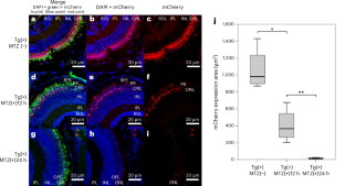
Zebrafish are widely used to investigate visual function, owing to their well-defined retinal organization and optical transparency during early developmental stages. Here, we evaluate the effectiveness of a color vision optomotor response (CV-OMR) assay for assessing zebrafish with red cone ablation. Tg (thrb:gal4;UAS:epNTR-p2a-mCherry) transgenic zebrafish were used to ablate red cones via the addition of metronidazole (MTZ). Transgenic zebrafish larvae were treated with MTZ for 0, 12 and 24 h at 5 days post-fertilization, resulting in Tg(+)MTZ(−), Tg(+)MTZ(+)12 h and Tg(+)MTZ(+)24 h groups, respectively. The areas of mCherry-expressing cells, representing red cones in tissue sections, were compared. The mean mCherry expression area was smallest in the Tg(+)MTZ(+)24 h group (16.5 ± 7.6 μm2), followed by the Tg(+)MTZ(+)12 h (404.1 ± 200.9 μm2) and Tg(+)MTZ(−) groups (1,066.6 ± 252.2 μm2; P < 0.001). At 6 days post-fertilization, zebrafish larvae were evaluated using the CV-OMR assay comprising two colors. Results were reported as the area under the curve of the ratio of larvae at the starting point curve. The ratio of larvae at the starting point decreased most rapidly in the Tg(+)MTZ(−) group and most slowly in the Tg(+)MTZ(+)24 h group. There were significant differences in the area under the curve of the ratio of larvae at the starting point curve among the three groups. In conclusion, the CV-OMR assay demonstrated the ability to differentiate color vision impairment based on the extent of red cone ablation. The CV-OMR assay used in this study may prove valuable for evaluating color vision in zebrafish with various eye diseases.
期刊介绍:
LabAnimal is a Nature Research journal dedicated to in vivo science and technology that improves our basic understanding and use of model organisms of human health and disease. In addition to basic research, methods and technologies, LabAnimal also covers important news, business and regulatory matters that impact the development and application of model organisms for preclinical research.
LabAnimal's focus is on innovative in vivo methods, research and technology covering a wide range of model organisms. Our broad scope ensures that the work we publish reaches the widest possible audience. LabAnimal provides a rigorous and fair peer review of manuscripts, high standards for copyediting and production, and efficient publication.

 求助内容:
求助内容: 应助结果提醒方式:


