{"title":"低增殖套细胞淋巴瘤患者继发性中枢神经系统淋巴瘤(SCNSL)的诊断。","authors":"Emmanuel Ojeabuo Oisakede, Ambreen Khalid, Chetan Patalappa, Faheem Anjum, Hashim Heyam","doi":"10.71480/nmj.v66i2.726","DOIUrl":null,"url":null,"abstract":"<p><p>Mantle cell lymphoma (MCL) represents a rare subtype of non-Hodgkin lymphoma, with approximately 4% of patients experiencing central nervous system (CNS) involvement. There is a higher likelihood of CNS Infiltration in patients who have elevated Ki-67 Levels, a marker associated with prognosis and CNS involvement. Herein, we present the case of a 72-year-old individual of British ancestry, known to have stage 4A MCL, exhibiting a Ki-67 index of 10%. He had just completed six cycles of Rituximab and Bendamustine with a good radiological response. The patient presented with episodic vacant states accompanied by confusion, recurrent dysphasia, and frontotemporal headaches followed by visual and auditory hallucinations upon hospital admission. Initial imaging studies failed to reveal compelling evidence of secondary central nervous system lymphoma (SCNSL), leading to a presumptive diagnosis of transient ischemic attack (TIA) given the patient's recurrent visits to the emergency department. However, subsequent evaluation by cerebrospinal fluid (CSF) analysis showed lymphocytic infiltrates with a negative bioFire test. Additional testing with the haematological malignancy diagnostic service (HMDS) unveiled findings consistent with CNS involvement secondary to known MCL. Our case report underscores the imperative for thorough CSF analysis in patients with MCL, irrespective of a seemingly low proliferation index such as Ki-67. This will promptly identify and manage potential CNS involvement.</p>","PeriodicalId":94346,"journal":{"name":"Nigerian medical journal : journal of the Nigeria Medical Association","volume":"66 2","pages":"805-810"},"PeriodicalIF":0.0000,"publicationDate":"2025-06-16","publicationTypes":"Journal Article","fieldsOfStudy":null,"isOpenAccess":false,"openAccessPdf":"https://www.ncbi.nlm.nih.gov/pmc/articles/PMC12280281/pdf/","citationCount":"0","resultStr":"{\"title\":\"Diagnosis of a secondary central nervous system Lymphoma (SCNSL) in a patient with low proliferative Mantle cell lymphoma.\",\"authors\":\"Emmanuel Ojeabuo Oisakede, Ambreen Khalid, Chetan Patalappa, Faheem Anjum, Hashim Heyam\",\"doi\":\"10.71480/nmj.v66i2.726\",\"DOIUrl\":null,\"url\":null,\"abstract\":\"<p><p>Mantle cell lymphoma (MCL) represents a rare subtype of non-Hodgkin lymphoma, with approximately 4% of patients experiencing central nervous system (CNS) involvement. There is a higher likelihood of CNS Infiltration in patients who have elevated Ki-67 Levels, a marker associated with prognosis and CNS involvement. Herein, we present the case of a 72-year-old individual of British ancestry, known to have stage 4A MCL, exhibiting a Ki-67 index of 10%. He had just completed six cycles of Rituximab and Bendamustine with a good radiological response. The patient presented with episodic vacant states accompanied by confusion, recurrent dysphasia, and frontotemporal headaches followed by visual and auditory hallucinations upon hospital admission. Initial imaging studies failed to reveal compelling evidence of secondary central nervous system lymphoma (SCNSL), leading to a presumptive diagnosis of transient ischemic attack (TIA) given the patient's recurrent visits to the emergency department. However, subsequent evaluation by cerebrospinal fluid (CSF) analysis showed lymphocytic infiltrates with a negative bioFire test. Additional testing with the haematological malignancy diagnostic service (HMDS) unveiled findings consistent with CNS involvement secondary to known MCL. Our case report underscores the imperative for thorough CSF analysis in patients with MCL, irrespective of a seemingly low proliferation index such as Ki-67. This will promptly identify and manage potential CNS involvement.</p>\",\"PeriodicalId\":94346,\"journal\":{\"name\":\"Nigerian medical journal : journal of the Nigeria Medical Association\",\"volume\":\"66 2\",\"pages\":\"805-810\"},\"PeriodicalIF\":0.0000,\"publicationDate\":\"2025-06-16\",\"publicationTypes\":\"Journal Article\",\"fieldsOfStudy\":null,\"isOpenAccess\":false,\"openAccessPdf\":\"https://www.ncbi.nlm.nih.gov/pmc/articles/PMC12280281/pdf/\",\"citationCount\":\"0\",\"resultStr\":null,\"platform\":\"Semanticscholar\",\"paperid\":null,\"PeriodicalName\":\"Nigerian medical journal : journal of the Nigeria Medical Association\",\"FirstCategoryId\":\"1085\",\"ListUrlMain\":\"https://doi.org/10.71480/nmj.v66i2.726\",\"RegionNum\":0,\"RegionCategory\":null,\"ArticlePicture\":[],\"TitleCN\":null,\"AbstractTextCN\":null,\"PMCID\":null,\"EPubDate\":\"2025/3/1 0:00:00\",\"PubModel\":\"eCollection\",\"JCR\":\"\",\"JCRName\":\"\",\"Score\":null,\"Total\":0}","platform":"Semanticscholar","paperid":null,"PeriodicalName":"Nigerian medical journal : journal of the Nigeria Medical Association","FirstCategoryId":"1085","ListUrlMain":"https://doi.org/10.71480/nmj.v66i2.726","RegionNum":0,"RegionCategory":null,"ArticlePicture":[],"TitleCN":null,"AbstractTextCN":null,"PMCID":null,"EPubDate":"2025/3/1 0:00:00","PubModel":"eCollection","JCR":"","JCRName":"","Score":null,"Total":0}
Diagnosis of a secondary central nervous system Lymphoma (SCNSL) in a patient with low proliferative Mantle cell lymphoma.
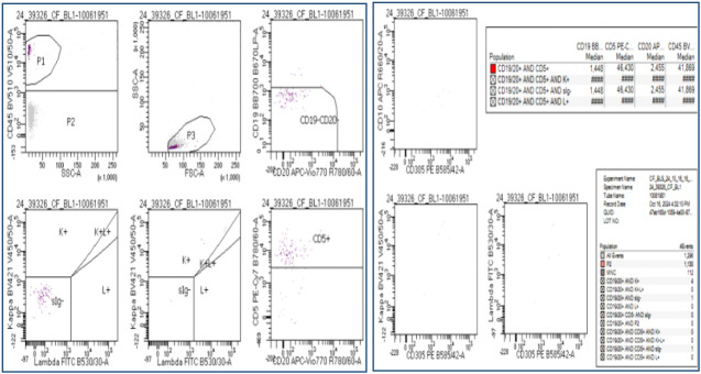
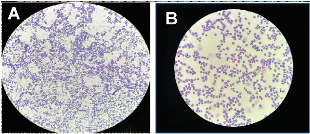
Mantle cell lymphoma (MCL) represents a rare subtype of non-Hodgkin lymphoma, with approximately 4% of patients experiencing central nervous system (CNS) involvement. There is a higher likelihood of CNS Infiltration in patients who have elevated Ki-67 Levels, a marker associated with prognosis and CNS involvement. Herein, we present the case of a 72-year-old individual of British ancestry, known to have stage 4A MCL, exhibiting a Ki-67 index of 10%. He had just completed six cycles of Rituximab and Bendamustine with a good radiological response. The patient presented with episodic vacant states accompanied by confusion, recurrent dysphasia, and frontotemporal headaches followed by visual and auditory hallucinations upon hospital admission. Initial imaging studies failed to reveal compelling evidence of secondary central nervous system lymphoma (SCNSL), leading to a presumptive diagnosis of transient ischemic attack (TIA) given the patient's recurrent visits to the emergency department. However, subsequent evaluation by cerebrospinal fluid (CSF) analysis showed lymphocytic infiltrates with a negative bioFire test. Additional testing with the haematological malignancy diagnostic service (HMDS) unveiled findings consistent with CNS involvement secondary to known MCL. Our case report underscores the imperative for thorough CSF analysis in patients with MCL, irrespective of a seemingly low proliferation index such as Ki-67. This will promptly identify and manage potential CNS involvement.


 求助内容:
求助内容: 应助结果提醒方式:


