{"title":"脉冲射频电流和热条件对人单核细胞β-内啡肽表达的影响。","authors":"Akira Nishioka, Toshiharu Azma, Tsutomu Mieda, Yasushi Mio","doi":"10.3390/neurosci6030067","DOIUrl":null,"url":null,"abstract":"<p><p>Pulsed radiofrequency (PRF) current applied to peripheral nerves is a modality used in interventional pain medicine, but its underlying mechanisms remain unclear. This study aimed to investigate whether ex vivo exposure of human monocytic THP-1 cells to PRF current or to heat induces β-endorphin production.</p><p><strong>Methods: </strong>THP-1 cells were exposed to PRF current for 15 min or incubated at elevated temperatures (42 °C to 50 °C) for 3 or 15 min. Flow cytometry was used to assess cell viability, and β-endorphin concentrations in culture supernatants were quantified by ELISA. In a separate experiment, cells were stimulated with lipopolysaccharide (LPS) to compare its effects on β-endorphin release.</p><p><strong>Results: </strong>A 3 min exposure to temperatures ≥ 46 °C reduced THP-1 cell viability, whereas a 15 min exposure to PRF current or to heat at 42 °C did not impair viability. Both PRF current and mild heat significantly enhanced β-endorphin release. β-Endorphin levels in the supernatant of LPS-stimulated cells were comparable to those of cells exposed to PRF current.</p><p><strong>Conclusions: </strong>Ex vivo application of PRF current or mild heat enhanced β-endorphin production from THP-1 cells without significant cytotoxicity. These preliminary findings warrant further investigation using primary human monocytes and in vivo models to assess therapeutic potential.</p>","PeriodicalId":74294,"journal":{"name":"NeuroSci","volume":"6 3","pages":""},"PeriodicalIF":2.0000,"publicationDate":"2025-07-21","publicationTypes":"Journal Article","fieldsOfStudy":null,"isOpenAccess":false,"openAccessPdf":"https://www.ncbi.nlm.nih.gov/pmc/articles/PMC12285971/pdf/","citationCount":"0","resultStr":"{\"title\":\"Effects of Pulsed Radiofrequency Current and Thermal Condition on the Expression of β-Endorphin in Human Monocytic Cells.\",\"authors\":\"Akira Nishioka, Toshiharu Azma, Tsutomu Mieda, Yasushi Mio\",\"doi\":\"10.3390/neurosci6030067\",\"DOIUrl\":null,\"url\":null,\"abstract\":\"<p><p>Pulsed radiofrequency (PRF) current applied to peripheral nerves is a modality used in interventional pain medicine, but its underlying mechanisms remain unclear. This study aimed to investigate whether ex vivo exposure of human monocytic THP-1 cells to PRF current or to heat induces β-endorphin production.</p><p><strong>Methods: </strong>THP-1 cells were exposed to PRF current for 15 min or incubated at elevated temperatures (42 °C to 50 °C) for 3 or 15 min. Flow cytometry was used to assess cell viability, and β-endorphin concentrations in culture supernatants were quantified by ELISA. In a separate experiment, cells were stimulated with lipopolysaccharide (LPS) to compare its effects on β-endorphin release.</p><p><strong>Results: </strong>A 3 min exposure to temperatures ≥ 46 °C reduced THP-1 cell viability, whereas a 15 min exposure to PRF current or to heat at 42 °C did not impair viability. Both PRF current and mild heat significantly enhanced β-endorphin release. β-Endorphin levels in the supernatant of LPS-stimulated cells were comparable to those of cells exposed to PRF current.</p><p><strong>Conclusions: </strong>Ex vivo application of PRF current or mild heat enhanced β-endorphin production from THP-1 cells without significant cytotoxicity. These preliminary findings warrant further investigation using primary human monocytes and in vivo models to assess therapeutic potential.</p>\",\"PeriodicalId\":74294,\"journal\":{\"name\":\"NeuroSci\",\"volume\":\"6 3\",\"pages\":\"\"},\"PeriodicalIF\":2.0000,\"publicationDate\":\"2025-07-21\",\"publicationTypes\":\"Journal Article\",\"fieldsOfStudy\":null,\"isOpenAccess\":false,\"openAccessPdf\":\"https://www.ncbi.nlm.nih.gov/pmc/articles/PMC12285971/pdf/\",\"citationCount\":\"0\",\"resultStr\":null,\"platform\":\"Semanticscholar\",\"paperid\":null,\"PeriodicalName\":\"NeuroSci\",\"FirstCategoryId\":\"1085\",\"ListUrlMain\":\"https://doi.org/10.3390/neurosci6030067\",\"RegionNum\":0,\"RegionCategory\":null,\"ArticlePicture\":[],\"TitleCN\":null,\"AbstractTextCN\":null,\"PMCID\":null,\"EPubDate\":\"\",\"PubModel\":\"\",\"JCR\":\"Q3\",\"JCRName\":\"CLINICAL NEUROLOGY\",\"Score\":null,\"Total\":0}","platform":"Semanticscholar","paperid":null,"PeriodicalName":"NeuroSci","FirstCategoryId":"1085","ListUrlMain":"https://doi.org/10.3390/neurosci6030067","RegionNum":0,"RegionCategory":null,"ArticlePicture":[],"TitleCN":null,"AbstractTextCN":null,"PMCID":null,"EPubDate":"","PubModel":"","JCR":"Q3","JCRName":"CLINICAL NEUROLOGY","Score":null,"Total":0}
Effects of Pulsed Radiofrequency Current and Thermal Condition on the Expression of β-Endorphin in Human Monocytic Cells.
Pulsed radiofrequency (PRF) current applied to peripheral nerves is a modality used in interventional pain medicine, but its underlying mechanisms remain unclear. This study aimed to investigate whether ex vivo exposure of human monocytic THP-1 cells to PRF current or to heat induces β-endorphin production.
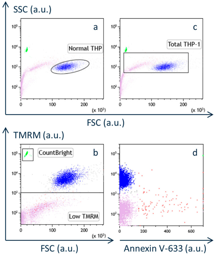
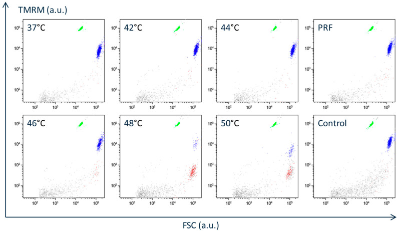
Methods: THP-1 cells were exposed to PRF current for 15 min or incubated at elevated temperatures (42 °C to 50 °C) for 3 or 15 min. Flow cytometry was used to assess cell viability, and β-endorphin concentrations in culture supernatants were quantified by ELISA. In a separate experiment, cells were stimulated with lipopolysaccharide (LPS) to compare its effects on β-endorphin release.
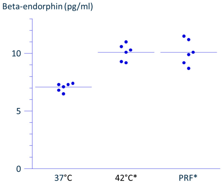
Results: A 3 min exposure to temperatures ≥ 46 °C reduced THP-1 cell viability, whereas a 15 min exposure to PRF current or to heat at 42 °C did not impair viability. Both PRF current and mild heat significantly enhanced β-endorphin release. β-Endorphin levels in the supernatant of LPS-stimulated cells were comparable to those of cells exposed to PRF current.
Conclusions: Ex vivo application of PRF current or mild heat enhanced β-endorphin production from THP-1 cells without significant cytotoxicity. These preliminary findings warrant further investigation using primary human monocytes and in vivo models to assess therapeutic potential.

 求助内容:
求助内容: 应助结果提醒方式:


