{"title":"阴道卵巢外勃勒纳瘤1例报告及文献复习。","authors":"Angel Yordanov, Milen Karaivanov, Stoyan Kostov, Vanya Savova, Vasilena Dimitrova","doi":"10.3390/reports8030103","DOIUrl":null,"url":null,"abstract":"<p><p><b>Background and Clinical Significance:</b> Brenner tumors are rare epithelial tumors that can occur in both males and females. They consist of ovarian transition cells surrounded by dense fibrous tissue and can be classified as benign, borderline, or malignant. While most commonly found in the ovary, extraovarian Brenner tumors (EOBTs) have been reported in the uterus, vagina, broad ligament, and omentum. <b>Case Presentation:</b> A 71-year-old postmenopausal woman presented with a polypous formation on the upper third of the posterior vaginal wall, which was found at a routine health check. Macroscopically, the lesion appeared as a solid, polypoid mass with a yellowish-gray cut surface, measuring approximately 25 × 20 mm. Histological examination revealed a polypoid formation covered by stratified squamous epithelium, with a dense fibrous stroma (Van Gieson [VG]+) and tubular structures lined by clear epithelial cells. Parenchymal cells showed low proliferative activity, with Ki-67 expression in less than 5% of cells, also Cytokeratin (CK) 7/+/p63:/+/ CK AE1/AE3: /+/ Estrogen Receptor (ER): /+/ and Progesterone Receptor (PR)/-/; CK20/-/; p53/-/, Wilms' Tumor (WT)-1/-/; Prostate-Specific Acid Phosphatase (PSAP)/-/. The final diagnosis was an extraovarian Brenner tumor. The patient was monitored for two months post-excision, with no signs of recurrence. <b>Conclusions:</b> EOBTs are extremely rarely seen and vaginal involvement is far less common. Due to their rarity, these tumors may be confused with other benign or malignant vaginal lesions. In order to differentiate EOBTs from other neoplasms, histological analysis is crucial due to their characteristic transitional-type epithelium and large fibrous stroma. Further studies are required to understand the origin and clinical behavior of EOBTs. Long-term monitoring should be performed to look for any recurrence or malignant change, even though benign Brenner tumors usually have a good prognosis. Awareness of EOBTs and their possible locations is essential for accurate diagnosis and appropriate management.</p>","PeriodicalId":74664,"journal":{"name":"Reports (MDPI)","volume":"8 3","pages":""},"PeriodicalIF":0.8000,"publicationDate":"2025-06-29","publicationTypes":"Journal Article","fieldsOfStudy":null,"isOpenAccess":false,"openAccessPdf":"https://www.ncbi.nlm.nih.gov/pmc/articles/PMC12265999/pdf/","citationCount":"0","resultStr":"{\"title\":\"Extraovarian Brenner Tumor in the Vagina: A Case Report and Review of Literature.\",\"authors\":\"Angel Yordanov, Milen Karaivanov, Stoyan Kostov, Vanya Savova, Vasilena Dimitrova\",\"doi\":\"10.3390/reports8030103\",\"DOIUrl\":null,\"url\":null,\"abstract\":\"<p><p><b>Background and Clinical Significance:</b> Brenner tumors are rare epithelial tumors that can occur in both males and females. They consist of ovarian transition cells surrounded by dense fibrous tissue and can be classified as benign, borderline, or malignant. While most commonly found in the ovary, extraovarian Brenner tumors (EOBTs) have been reported in the uterus, vagina, broad ligament, and omentum. <b>Case Presentation:</b> A 71-year-old postmenopausal woman presented with a polypous formation on the upper third of the posterior vaginal wall, which was found at a routine health check. Macroscopically, the lesion appeared as a solid, polypoid mass with a yellowish-gray cut surface, measuring approximately 25 × 20 mm. Histological examination revealed a polypoid formation covered by stratified squamous epithelium, with a dense fibrous stroma (Van Gieson [VG]+) and tubular structures lined by clear epithelial cells. Parenchymal cells showed low proliferative activity, with Ki-67 expression in less than 5% of cells, also Cytokeratin (CK) 7/+/p63:/+/ CK AE1/AE3: /+/ Estrogen Receptor (ER): /+/ and Progesterone Receptor (PR)/-/; CK20/-/; p53/-/, Wilms' Tumor (WT)-1/-/; Prostate-Specific Acid Phosphatase (PSAP)/-/. The final diagnosis was an extraovarian Brenner tumor. The patient was monitored for two months post-excision, with no signs of recurrence. <b>Conclusions:</b> EOBTs are extremely rarely seen and vaginal involvement is far less common. Due to their rarity, these tumors may be confused with other benign or malignant vaginal lesions. In order to differentiate EOBTs from other neoplasms, histological analysis is crucial due to their characteristic transitional-type epithelium and large fibrous stroma. Further studies are required to understand the origin and clinical behavior of EOBTs. Long-term monitoring should be performed to look for any recurrence or malignant change, even though benign Brenner tumors usually have a good prognosis. Awareness of EOBTs and their possible locations is essential for accurate diagnosis and appropriate management.</p>\",\"PeriodicalId\":74664,\"journal\":{\"name\":\"Reports (MDPI)\",\"volume\":\"8 3\",\"pages\":\"\"},\"PeriodicalIF\":0.8000,\"publicationDate\":\"2025-06-29\",\"publicationTypes\":\"Journal Article\",\"fieldsOfStudy\":null,\"isOpenAccess\":false,\"openAccessPdf\":\"https://www.ncbi.nlm.nih.gov/pmc/articles/PMC12265999/pdf/\",\"citationCount\":\"0\",\"resultStr\":null,\"platform\":\"Semanticscholar\",\"paperid\":null,\"PeriodicalName\":\"Reports (MDPI)\",\"FirstCategoryId\":\"1085\",\"ListUrlMain\":\"https://doi.org/10.3390/reports8030103\",\"RegionNum\":0,\"RegionCategory\":null,\"ArticlePicture\":[],\"TitleCN\":null,\"AbstractTextCN\":null,\"PMCID\":null,\"EPubDate\":\"\",\"PubModel\":\"\",\"JCR\":\"Q3\",\"JCRName\":\"MEDICINE, GENERAL & INTERNAL\",\"Score\":null,\"Total\":0}","platform":"Semanticscholar","paperid":null,"PeriodicalName":"Reports (MDPI)","FirstCategoryId":"1085","ListUrlMain":"https://doi.org/10.3390/reports8030103","RegionNum":0,"RegionCategory":null,"ArticlePicture":[],"TitleCN":null,"AbstractTextCN":null,"PMCID":null,"EPubDate":"","PubModel":"","JCR":"Q3","JCRName":"MEDICINE, GENERAL & INTERNAL","Score":null,"Total":0}
Extraovarian Brenner Tumor in the Vagina: A Case Report and Review of Literature.
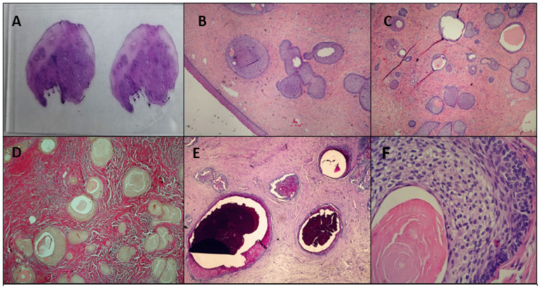
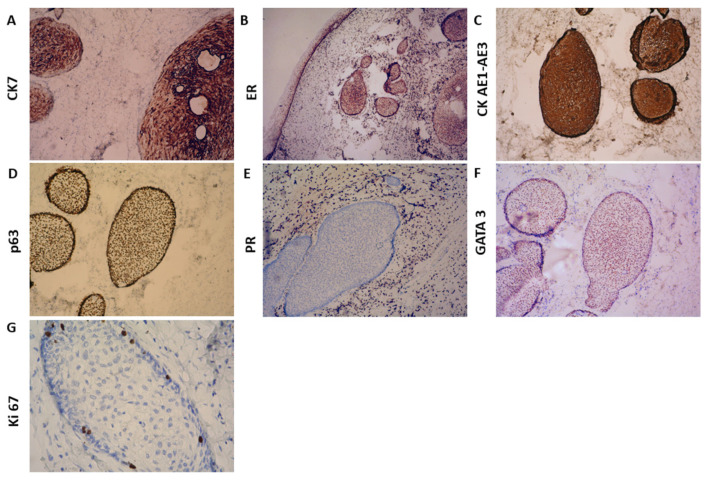
Background and Clinical Significance: Brenner tumors are rare epithelial tumors that can occur in both males and females. They consist of ovarian transition cells surrounded by dense fibrous tissue and can be classified as benign, borderline, or malignant. While most commonly found in the ovary, extraovarian Brenner tumors (EOBTs) have been reported in the uterus, vagina, broad ligament, and omentum. Case Presentation: A 71-year-old postmenopausal woman presented with a polypous formation on the upper third of the posterior vaginal wall, which was found at a routine health check. Macroscopically, the lesion appeared as a solid, polypoid mass with a yellowish-gray cut surface, measuring approximately 25 × 20 mm. Histological examination revealed a polypoid formation covered by stratified squamous epithelium, with a dense fibrous stroma (Van Gieson [VG]+) and tubular structures lined by clear epithelial cells. Parenchymal cells showed low proliferative activity, with Ki-67 expression in less than 5% of cells, also Cytokeratin (CK) 7/+/p63:/+/ CK AE1/AE3: /+/ Estrogen Receptor (ER): /+/ and Progesterone Receptor (PR)/-/; CK20/-/; p53/-/, Wilms' Tumor (WT)-1/-/; Prostate-Specific Acid Phosphatase (PSAP)/-/. The final diagnosis was an extraovarian Brenner tumor. The patient was monitored for two months post-excision, with no signs of recurrence. Conclusions: EOBTs are extremely rarely seen and vaginal involvement is far less common. Due to their rarity, these tumors may be confused with other benign or malignant vaginal lesions. In order to differentiate EOBTs from other neoplasms, histological analysis is crucial due to their characteristic transitional-type epithelium and large fibrous stroma. Further studies are required to understand the origin and clinical behavior of EOBTs. Long-term monitoring should be performed to look for any recurrence or malignant change, even though benign Brenner tumors usually have a good prognosis. Awareness of EOBTs and their possible locations is essential for accurate diagnosis and appropriate management.

 求助内容:
求助内容: 应助结果提醒方式:


