{"title":"老年人肩关节疾病的影像学特征,包括粘连性囊炎、肩袖撕裂和肩关节骨关节炎:叙述性回顾","authors":"Myung-Seo Kim, Tae-Hoon Jung","doi":"10.12771/emj.2025.e10","DOIUrl":null,"url":null,"abstract":"<p><p>Shoulder diseases, including adhesive capsulitis, rotator cuff tear, and osteoarthritis of the glenohumeral joint, can significantly impair daily activities in older adult patients. This review aims to examine the radiologic findings associated with these shoulder conditions in older patients, providing insights for accurate diagnosis and effective treatment. Adhesive capsulitis, commonly known as frozen shoulder, leads to pain and restricted movement, thereby causing shoulder dysfunction. Recent advances in diagnostic technology have greatly enhanced the sensitivity and accuracy of diagnosing this condition through radiologic evaluations, including MRI, magnetic resonance arthrography (MRA), and high-resolution ultrasound. Rotator cuff disease is another frequent issue in older adults, with full-thickness tears occurring in 50%-80% of cases. Both MRI and MRA are highly sensitive and specific in identifying rotator cuff tears. Additionally, ultrasonography is recognized for its high sensitivity and specificity in detecting tears of the supraspinatus tendon. Although osteoarthritis of the glenohumeral joint is less commonly prevalent, its advanced stages can severely affect the function of the upper extremity. Plain radiography is typically the first imaging technique used to assess this type of osteoarthritis. As the condition worsens, CT is utilized to measure glenoid bone loss, glenoid version, and inclination, which are crucial for accurate surgical planning. Each imaging modality provides distinct benefits: plain radiographs for initial structural assessment, ultrasonography for real-time evaluation of soft tissues, MRI/MRA for detailed visualization of capsular and tendinous lesions, and CT for precise bony analysis.</p>","PeriodicalId":41392,"journal":{"name":"Ewha Medical Journal","volume":"48 1","pages":"e10"},"PeriodicalIF":0.2000,"publicationDate":"2025-01-01","publicationTypes":"Journal Article","fieldsOfStudy":null,"isOpenAccess":false,"openAccessPdf":"https://www.ncbi.nlm.nih.gov/pmc/articles/PMC12277897/pdf/","citationCount":"0","resultStr":"{\"title\":\"Radiological characteristics of shoulder diseases in older adults, including adhesive capsulitis, rotator cuff tear, and osteoarthritis of the glenohumeral joint: a narrative review.\",\"authors\":\"Myung-Seo Kim, Tae-Hoon Jung\",\"doi\":\"10.12771/emj.2025.e10\",\"DOIUrl\":null,\"url\":null,\"abstract\":\"<p><p>Shoulder diseases, including adhesive capsulitis, rotator cuff tear, and osteoarthritis of the glenohumeral joint, can significantly impair daily activities in older adult patients. This review aims to examine the radiologic findings associated with these shoulder conditions in older patients, providing insights for accurate diagnosis and effective treatment. Adhesive capsulitis, commonly known as frozen shoulder, leads to pain and restricted movement, thereby causing shoulder dysfunction. Recent advances in diagnostic technology have greatly enhanced the sensitivity and accuracy of diagnosing this condition through radiologic evaluations, including MRI, magnetic resonance arthrography (MRA), and high-resolution ultrasound. Rotator cuff disease is another frequent issue in older adults, with full-thickness tears occurring in 50%-80% of cases. Both MRI and MRA are highly sensitive and specific in identifying rotator cuff tears. Additionally, ultrasonography is recognized for its high sensitivity and specificity in detecting tears of the supraspinatus tendon. Although osteoarthritis of the glenohumeral joint is less commonly prevalent, its advanced stages can severely affect the function of the upper extremity. Plain radiography is typically the first imaging technique used to assess this type of osteoarthritis. As the condition worsens, CT is utilized to measure glenoid bone loss, glenoid version, and inclination, which are crucial for accurate surgical planning. Each imaging modality provides distinct benefits: plain radiographs for initial structural assessment, ultrasonography for real-time evaluation of soft tissues, MRI/MRA for detailed visualization of capsular and tendinous lesions, and CT for precise bony analysis.</p>\",\"PeriodicalId\":41392,\"journal\":{\"name\":\"Ewha Medical Journal\",\"volume\":\"48 1\",\"pages\":\"e10\"},\"PeriodicalIF\":0.2000,\"publicationDate\":\"2025-01-01\",\"publicationTypes\":\"Journal Article\",\"fieldsOfStudy\":null,\"isOpenAccess\":false,\"openAccessPdf\":\"https://www.ncbi.nlm.nih.gov/pmc/articles/PMC12277897/pdf/\",\"citationCount\":\"0\",\"resultStr\":null,\"platform\":\"Semanticscholar\",\"paperid\":null,\"PeriodicalName\":\"Ewha Medical Journal\",\"FirstCategoryId\":\"1085\",\"ListUrlMain\":\"https://doi.org/10.12771/emj.2025.e10\",\"RegionNum\":0,\"RegionCategory\":null,\"ArticlePicture\":[],\"TitleCN\":null,\"AbstractTextCN\":null,\"PMCID\":null,\"EPubDate\":\"2025/1/31 0:00:00\",\"PubModel\":\"Epub\",\"JCR\":\"Q3\",\"JCRName\":\"MEDICINE, GENERAL & INTERNAL\",\"Score\":null,\"Total\":0}","platform":"Semanticscholar","paperid":null,"PeriodicalName":"Ewha Medical Journal","FirstCategoryId":"1085","ListUrlMain":"https://doi.org/10.12771/emj.2025.e10","RegionNum":0,"RegionCategory":null,"ArticlePicture":[],"TitleCN":null,"AbstractTextCN":null,"PMCID":null,"EPubDate":"2025/1/31 0:00:00","PubModel":"Epub","JCR":"Q3","JCRName":"MEDICINE, GENERAL & INTERNAL","Score":null,"Total":0}
Radiological characteristics of shoulder diseases in older adults, including adhesive capsulitis, rotator cuff tear, and osteoarthritis of the glenohumeral joint: a narrative review.
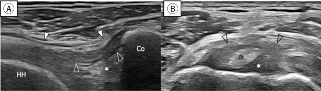
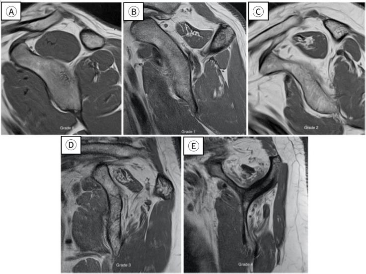
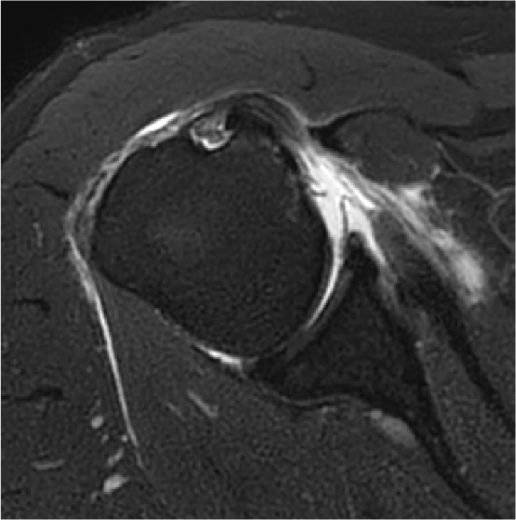
Shoulder diseases, including adhesive capsulitis, rotator cuff tear, and osteoarthritis of the glenohumeral joint, can significantly impair daily activities in older adult patients. This review aims to examine the radiologic findings associated with these shoulder conditions in older patients, providing insights for accurate diagnosis and effective treatment. Adhesive capsulitis, commonly known as frozen shoulder, leads to pain and restricted movement, thereby causing shoulder dysfunction. Recent advances in diagnostic technology have greatly enhanced the sensitivity and accuracy of diagnosing this condition through radiologic evaluations, including MRI, magnetic resonance arthrography (MRA), and high-resolution ultrasound. Rotator cuff disease is another frequent issue in older adults, with full-thickness tears occurring in 50%-80% of cases. Both MRI and MRA are highly sensitive and specific in identifying rotator cuff tears. Additionally, ultrasonography is recognized for its high sensitivity and specificity in detecting tears of the supraspinatus tendon. Although osteoarthritis of the glenohumeral joint is less commonly prevalent, its advanced stages can severely affect the function of the upper extremity. Plain radiography is typically the first imaging technique used to assess this type of osteoarthritis. As the condition worsens, CT is utilized to measure glenoid bone loss, glenoid version, and inclination, which are crucial for accurate surgical planning. Each imaging modality provides distinct benefits: plain radiographs for initial structural assessment, ultrasonography for real-time evaluation of soft tissues, MRI/MRA for detailed visualization of capsular and tendinous lesions, and CT for precise bony analysis.

 求助内容:
求助内容: 应助结果提醒方式:


