{"title":"SOX2和OCT4在原发性牙源性角化囊肿、复发性牙源性角化囊肿和减压术治疗牙源性角化囊肿中的免疫组化表达。","authors":"Chinmayee Mannava, Ravikanth Manyam, Nimmagadda Vikas Kumar, Divya Naga Lakshmi Puvvada, P Swetha, Naga Supriya","doi":"10.4103/jomfp.jomfp_254_24","DOIUrl":null,"url":null,"abstract":"<p><strong>Introduction: </strong>Odontogenic keratocyst (OKC) is a developmental odontogenic cyst with distinct pathological features and a high recurrence rate. Interest among OKCs became apparent by the clinical challenges associated with their treatment. Pathogenesis of OKCs is a multifactorial process, which is linked to several signalling pathways and expression of stem cell markers such as SOX2 and OCT4.</p><p><strong>Materials and methods: </strong>Thirty cases of OKCs were categorised into three groups: primary (n = 10), recurrent (n = 10), and decompressed (n = 10). Tissue sections were immunohistochemically stained using anti-SOX2 and anti-OCT4 antibodies. Staining distribution, intensity, and localisation were evaluated qualitatively. Quantitative assessment was performed using Image Pro Plus software, and statistical analysis was conducted using SPSS software and results were statistically analysed.</p><p><strong>Results: </strong>SOX2 expression was observed in 80% of primary, 80% recurrent, and 90% of decompressed OKCs, with significant differences in staining intensity (<i>P</i> = 0.032). Most cases exhibited diffuse, nuclear, and cytoplasmic positivity across the full epithelial thickness, particularly in suprabasal layers. OCT4 expression was limited to 10% of primary and 20% of recurrent OKCs, with no positivity observed in decompressed cases. OCT4 did not show statistically significant differences. Remmele scores for both markers were not statistically significant across the groups.</p><p><strong>Conclusion: </strong>High expression of SOX2 in OKCs supports its role as a marker of epithelial stemness and a potential biomarker for aggressive behaviour and recurrence. Limited expression of OCT4 suggests a minimal role in OKC pathobiology, possibly associated with early differentiation. Lack of OCT4 expression in decompressed lesions raises questions about the molecular efficacy of decompression therapy.</p>","PeriodicalId":38846,"journal":{"name":"Journal of Oral and Maxillofacial Pathology","volume":"29 2","pages":"286-292"},"PeriodicalIF":0.0000,"publicationDate":"2025-04-01","publicationTypes":"Journal Article","fieldsOfStudy":null,"isOpenAccess":false,"openAccessPdf":"https://www.ncbi.nlm.nih.gov/pmc/articles/PMC12283042/pdf/","citationCount":"0","resultStr":"{\"title\":\"Immunohistochemical expression of SOX2 and OCT4 in primary odontogenic keratocyst, recurrent odontogenic keratocyst, and odontogenic keratocyst treated by the decompression technique.\",\"authors\":\"Chinmayee Mannava, Ravikanth Manyam, Nimmagadda Vikas Kumar, Divya Naga Lakshmi Puvvada, P Swetha, Naga Supriya\",\"doi\":\"10.4103/jomfp.jomfp_254_24\",\"DOIUrl\":null,\"url\":null,\"abstract\":\"<p><strong>Introduction: </strong>Odontogenic keratocyst (OKC) is a developmental odontogenic cyst with distinct pathological features and a high recurrence rate. Interest among OKCs became apparent by the clinical challenges associated with their treatment. Pathogenesis of OKCs is a multifactorial process, which is linked to several signalling pathways and expression of stem cell markers such as SOX2 and OCT4.</p><p><strong>Materials and methods: </strong>Thirty cases of OKCs were categorised into three groups: primary (n = 10), recurrent (n = 10), and decompressed (n = 10). Tissue sections were immunohistochemically stained using anti-SOX2 and anti-OCT4 antibodies. Staining distribution, intensity, and localisation were evaluated qualitatively. Quantitative assessment was performed using Image Pro Plus software, and statistical analysis was conducted using SPSS software and results were statistically analysed.</p><p><strong>Results: </strong>SOX2 expression was observed in 80% of primary, 80% recurrent, and 90% of decompressed OKCs, with significant differences in staining intensity (<i>P</i> = 0.032). Most cases exhibited diffuse, nuclear, and cytoplasmic positivity across the full epithelial thickness, particularly in suprabasal layers. OCT4 expression was limited to 10% of primary and 20% of recurrent OKCs, with no positivity observed in decompressed cases. OCT4 did not show statistically significant differences. Remmele scores for both markers were not statistically significant across the groups.</p><p><strong>Conclusion: </strong>High expression of SOX2 in OKCs supports its role as a marker of epithelial stemness and a potential biomarker for aggressive behaviour and recurrence. Limited expression of OCT4 suggests a minimal role in OKC pathobiology, possibly associated with early differentiation. Lack of OCT4 expression in decompressed lesions raises questions about the molecular efficacy of decompression therapy.</p>\",\"PeriodicalId\":38846,\"journal\":{\"name\":\"Journal of Oral and Maxillofacial Pathology\",\"volume\":\"29 2\",\"pages\":\"286-292\"},\"PeriodicalIF\":0.0000,\"publicationDate\":\"2025-04-01\",\"publicationTypes\":\"Journal Article\",\"fieldsOfStudy\":null,\"isOpenAccess\":false,\"openAccessPdf\":\"https://www.ncbi.nlm.nih.gov/pmc/articles/PMC12283042/pdf/\",\"citationCount\":\"0\",\"resultStr\":null,\"platform\":\"Semanticscholar\",\"paperid\":null,\"PeriodicalName\":\"Journal of Oral and Maxillofacial Pathology\",\"FirstCategoryId\":\"1085\",\"ListUrlMain\":\"https://doi.org/10.4103/jomfp.jomfp_254_24\",\"RegionNum\":0,\"RegionCategory\":null,\"ArticlePicture\":[],\"TitleCN\":null,\"AbstractTextCN\":null,\"PMCID\":null,\"EPubDate\":\"2025/6/30 0:00:00\",\"PubModel\":\"Epub\",\"JCR\":\"Q3\",\"JCRName\":\"Medicine\",\"Score\":null,\"Total\":0}","platform":"Semanticscholar","paperid":null,"PeriodicalName":"Journal of Oral and Maxillofacial Pathology","FirstCategoryId":"1085","ListUrlMain":"https://doi.org/10.4103/jomfp.jomfp_254_24","RegionNum":0,"RegionCategory":null,"ArticlePicture":[],"TitleCN":null,"AbstractTextCN":null,"PMCID":null,"EPubDate":"2025/6/30 0:00:00","PubModel":"Epub","JCR":"Q3","JCRName":"Medicine","Score":null,"Total":0}
Immunohistochemical expression of SOX2 and OCT4 in primary odontogenic keratocyst, recurrent odontogenic keratocyst, and odontogenic keratocyst treated by the decompression technique.
Introduction: Odontogenic keratocyst (OKC) is a developmental odontogenic cyst with distinct pathological features and a high recurrence rate. Interest among OKCs became apparent by the clinical challenges associated with their treatment. Pathogenesis of OKCs is a multifactorial process, which is linked to several signalling pathways and expression of stem cell markers such as SOX2 and OCT4.
Materials and methods: Thirty cases of OKCs were categorised into three groups: primary (n = 10), recurrent (n = 10), and decompressed (n = 10). Tissue sections were immunohistochemically stained using anti-SOX2 and anti-OCT4 antibodies. Staining distribution, intensity, and localisation were evaluated qualitatively. Quantitative assessment was performed using Image Pro Plus software, and statistical analysis was conducted using SPSS software and results were statistically analysed.
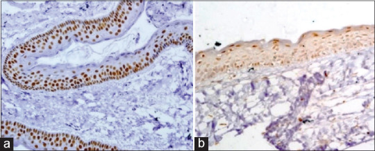
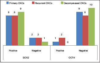
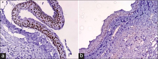
Results: SOX2 expression was observed in 80% of primary, 80% recurrent, and 90% of decompressed OKCs, with significant differences in staining intensity (P = 0.032). Most cases exhibited diffuse, nuclear, and cytoplasmic positivity across the full epithelial thickness, particularly in suprabasal layers. OCT4 expression was limited to 10% of primary and 20% of recurrent OKCs, with no positivity observed in decompressed cases. OCT4 did not show statistically significant differences. Remmele scores for both markers were not statistically significant across the groups.
Conclusion: High expression of SOX2 in OKCs supports its role as a marker of epithelial stemness and a potential biomarker for aggressive behaviour and recurrence. Limited expression of OCT4 suggests a minimal role in OKC pathobiology, possibly associated with early differentiation. Lack of OCT4 expression in decompressed lesions raises questions about the molecular efficacy of decompression therapy.
期刊介绍:
The journal of Oral and Maxillofacial Pathology [ISSN:print-(0973-029X, online-1998-393X)] is a tri-annual journal published on behalf of “The Indian Association of Oral and Maxillofacial Pathologists” (IAOMP). The publication of JOMFP was started in the year 1993. The journal publishes papers on a wide spectrum of topics associated with the scope of Oral and Maxillofacial Pathology, also, ensuring scientific merit and quality. It is a comprehensive reading material for the professionals who want to upgrade their diagnostic skills in Oral Diseases; allows exposure to newer topics and methods of research in the Oral-facial Tissues and Pathology. New features allow an open minded thinking and approach to various pathologies. It also encourages authors to showcase quality work done by them and to compile relevant cases which are diagnostically challenging. The Journal takes pride in maintaining the quality of articles and photomicrographs.

 求助内容:
求助内容: 应助结果提醒方式:


