{"title":"壳聚糖纳米颗粒经黏膜递送miR-30c-5p治疗口腔鳞状细胞癌","authors":"Yi-Ping Fang, Yu-Chih Lin, Chien-Yu Lin, Po-Jen Wang, Ting-Yu Chang, Ya-Ju Hsieh","doi":"10.2147/IJN.S524558","DOIUrl":null,"url":null,"abstract":"<p><strong>Background: </strong>Oral squamous cell carcinoma (OSCC) remains difficult to treat with current modalities. miR-30c-5p, a tumor-suppressive microRNA frequently downregulated in OSCC, inhibits cancer cell proliferation and migration. However, its clinical application is limited by poor stability and inefficient uptake. To address these issues, miR-30c-5p was encapsulated into chitosan nanoparticles (CS-NPs) using ionic gelation to enhance delivery and protect against degradation.</p><p><strong>Methods: </strong>miR-30c-5p-loaded CS-NPs were characterized for particle size, zeta potential, morphology, and encapsulation efficiency. HSC-3 and OEC-M1 cells were treated with free miRNA, CS-NPs, or CS-miRNA-NPs at final concentrations of 5%, 10%, 25%, and 50% (v/v) in culture medium. Cellular uptake was assessed by confocal microscopy. Ex vivo porcine buccal membrane studies evaluated mucosal penetration. Cytotoxicity was determined using MTT assays, while gene regulation was analyzed via quantitative polymerase chain reaction and Western blotting.</p><p><strong>Results: </strong>The prepared CS-NPs had particle sizes ranging from 434 to 452 nm and encapsulation efficiencies between 79% and 87%. Confocal imaging revealed significantly greater cytoplasmic uptake of CS-miRNA-FAM NPs versus free miRNA. Ex vivo studies showed that CS-miR-30c-5p-FAM NPs penetrated mucosa up to 80-160 μm with a 5.42-fold higher fluorescence intensity than free miR-30c-5p-FAM. Cytotoxicity testing showed high cell viability (>93%) for all treatments at concentrations ≤25% (v/v). At 50% (v/v) nanoparticle suspension, viability significantly decreased in OEC-M1 cells (84.41% for naked miRNA, 54.52% for CS-NPs, 61.10% for CS-miRNA-NPs; P < 0.001). After 48 h, greater reductions were observed at 50% (v/v), with cell viability in HSC-3 cells decreasing to 85.55% (free miRNA), 42.72% (CS-NPs), and 51.82% (CS-miRNA-NPs), and in OEC-M1 cells to 73.98%, 33.00%, and 39.89%, respectively. Functional assays showed vimentin mRNA reductions of 85% in HSC-3 and 30% in OEC-M1, with protein decreases confirmed by Western blot.</p><p><strong>Conclusion: </strong>CS-NPs enhance miRNA delivery and gene-silencing efficacy in OSCC cells. These findings support CS-based systems for miRNA therapeutics in oral cancer.</p>","PeriodicalId":14084,"journal":{"name":"International Journal of Nanomedicine","volume":"20 ","pages":"9179-9194"},"PeriodicalIF":6.5000,"publicationDate":"2025-07-19","publicationTypes":"Journal Article","fieldsOfStudy":null,"isOpenAccess":false,"openAccessPdf":"https://www.ncbi.nlm.nih.gov/pmc/articles/PMC12285890/pdf/","citationCount":"0","resultStr":"{\"title\":\"Transmucosal Delivery of miR-30c-5p by Chitosan Nanoparticles for Oral Squamous Cell Carcinoma.\",\"authors\":\"Yi-Ping Fang, Yu-Chih Lin, Chien-Yu Lin, Po-Jen Wang, Ting-Yu Chang, Ya-Ju Hsieh\",\"doi\":\"10.2147/IJN.S524558\",\"DOIUrl\":null,\"url\":null,\"abstract\":\"<p><strong>Background: </strong>Oral squamous cell carcinoma (OSCC) remains difficult to treat with current modalities. miR-30c-5p, a tumor-suppressive microRNA frequently downregulated in OSCC, inhibits cancer cell proliferation and migration. However, its clinical application is limited by poor stability and inefficient uptake. To address these issues, miR-30c-5p was encapsulated into chitosan nanoparticles (CS-NPs) using ionic gelation to enhance delivery and protect against degradation.</p><p><strong>Methods: </strong>miR-30c-5p-loaded CS-NPs were characterized for particle size, zeta potential, morphology, and encapsulation efficiency. HSC-3 and OEC-M1 cells were treated with free miRNA, CS-NPs, or CS-miRNA-NPs at final concentrations of 5%, 10%, 25%, and 50% (v/v) in culture medium. Cellular uptake was assessed by confocal microscopy. Ex vivo porcine buccal membrane studies evaluated mucosal penetration. Cytotoxicity was determined using MTT assays, while gene regulation was analyzed via quantitative polymerase chain reaction and Western blotting.</p><p><strong>Results: </strong>The prepared CS-NPs had particle sizes ranging from 434 to 452 nm and encapsulation efficiencies between 79% and 87%. Confocal imaging revealed significantly greater cytoplasmic uptake of CS-miRNA-FAM NPs versus free miRNA. Ex vivo studies showed that CS-miR-30c-5p-FAM NPs penetrated mucosa up to 80-160 μm with a 5.42-fold higher fluorescence intensity than free miR-30c-5p-FAM. Cytotoxicity testing showed high cell viability (>93%) for all treatments at concentrations ≤25% (v/v). At 50% (v/v) nanoparticle suspension, viability significantly decreased in OEC-M1 cells (84.41% for naked miRNA, 54.52% for CS-NPs, 61.10% for CS-miRNA-NPs; P < 0.001). After 48 h, greater reductions were observed at 50% (v/v), with cell viability in HSC-3 cells decreasing to 85.55% (free miRNA), 42.72% (CS-NPs), and 51.82% (CS-miRNA-NPs), and in OEC-M1 cells to 73.98%, 33.00%, and 39.89%, respectively. Functional assays showed vimentin mRNA reductions of 85% in HSC-3 and 30% in OEC-M1, with protein decreases confirmed by Western blot.</p><p><strong>Conclusion: </strong>CS-NPs enhance miRNA delivery and gene-silencing efficacy in OSCC cells. These findings support CS-based systems for miRNA therapeutics in oral cancer.</p>\",\"PeriodicalId\":14084,\"journal\":{\"name\":\"International Journal of Nanomedicine\",\"volume\":\"20 \",\"pages\":\"9179-9194\"},\"PeriodicalIF\":6.5000,\"publicationDate\":\"2025-07-19\",\"publicationTypes\":\"Journal Article\",\"fieldsOfStudy\":null,\"isOpenAccess\":false,\"openAccessPdf\":\"https://www.ncbi.nlm.nih.gov/pmc/articles/PMC12285890/pdf/\",\"citationCount\":\"0\",\"resultStr\":null,\"platform\":\"Semanticscholar\",\"paperid\":null,\"PeriodicalName\":\"International Journal of Nanomedicine\",\"FirstCategoryId\":\"3\",\"ListUrlMain\":\"https://doi.org/10.2147/IJN.S524558\",\"RegionNum\":2,\"RegionCategory\":\"医学\",\"ArticlePicture\":[],\"TitleCN\":null,\"AbstractTextCN\":null,\"PMCID\":null,\"EPubDate\":\"2025/1/1 0:00:00\",\"PubModel\":\"eCollection\",\"JCR\":\"Q1\",\"JCRName\":\"NANOSCIENCE & NANOTECHNOLOGY\",\"Score\":null,\"Total\":0}","platform":"Semanticscholar","paperid":null,"PeriodicalName":"International Journal of Nanomedicine","FirstCategoryId":"3","ListUrlMain":"https://doi.org/10.2147/IJN.S524558","RegionNum":2,"RegionCategory":"医学","ArticlePicture":[],"TitleCN":null,"AbstractTextCN":null,"PMCID":null,"EPubDate":"2025/1/1 0:00:00","PubModel":"eCollection","JCR":"Q1","JCRName":"NANOSCIENCE & NANOTECHNOLOGY","Score":null,"Total":0}
Transmucosal Delivery of miR-30c-5p by Chitosan Nanoparticles for Oral Squamous Cell Carcinoma.
Background: Oral squamous cell carcinoma (OSCC) remains difficult to treat with current modalities. miR-30c-5p, a tumor-suppressive microRNA frequently downregulated in OSCC, inhibits cancer cell proliferation and migration. However, its clinical application is limited by poor stability and inefficient uptake. To address these issues, miR-30c-5p was encapsulated into chitosan nanoparticles (CS-NPs) using ionic gelation to enhance delivery and protect against degradation.
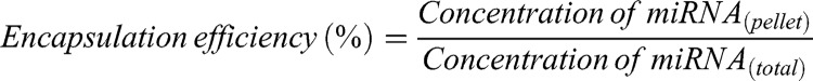
Methods: miR-30c-5p-loaded CS-NPs were characterized for particle size, zeta potential, morphology, and encapsulation efficiency. HSC-3 and OEC-M1 cells were treated with free miRNA, CS-NPs, or CS-miRNA-NPs at final concentrations of 5%, 10%, 25%, and 50% (v/v) in culture medium. Cellular uptake was assessed by confocal microscopy. Ex vivo porcine buccal membrane studies evaluated mucosal penetration. Cytotoxicity was determined using MTT assays, while gene regulation was analyzed via quantitative polymerase chain reaction and Western blotting.
Results: The prepared CS-NPs had particle sizes ranging from 434 to 452 nm and encapsulation efficiencies between 79% and 87%. Confocal imaging revealed significantly greater cytoplasmic uptake of CS-miRNA-FAM NPs versus free miRNA. Ex vivo studies showed that CS-miR-30c-5p-FAM NPs penetrated mucosa up to 80-160 μm with a 5.42-fold higher fluorescence intensity than free miR-30c-5p-FAM. Cytotoxicity testing showed high cell viability (>93%) for all treatments at concentrations ≤25% (v/v). At 50% (v/v) nanoparticle suspension, viability significantly decreased in OEC-M1 cells (84.41% for naked miRNA, 54.52% for CS-NPs, 61.10% for CS-miRNA-NPs; P < 0.001). After 48 h, greater reductions were observed at 50% (v/v), with cell viability in HSC-3 cells decreasing to 85.55% (free miRNA), 42.72% (CS-NPs), and 51.82% (CS-miRNA-NPs), and in OEC-M1 cells to 73.98%, 33.00%, and 39.89%, respectively. Functional assays showed vimentin mRNA reductions of 85% in HSC-3 and 30% in OEC-M1, with protein decreases confirmed by Western blot.
Conclusion: CS-NPs enhance miRNA delivery and gene-silencing efficacy in OSCC cells. These findings support CS-based systems for miRNA therapeutics in oral cancer.
期刊介绍:
The International Journal of Nanomedicine is a globally recognized journal that focuses on the applications of nanotechnology in the biomedical field. It is a peer-reviewed and open-access publication that covers diverse aspects of this rapidly evolving research area.
With its strong emphasis on the clinical potential of nanoparticles in disease diagnostics, prevention, and treatment, the journal aims to showcase cutting-edge research and development in the field.
Starting from now, the International Journal of Nanomedicine will not accept meta-analyses for publication.


 求助内容:
求助内容: 应助结果提醒方式:


