{"title":"神经节细胞层厚度作为肌萎缩侧索硬化功能结局的生物标志物:一项OCT研究。","authors":"Divya Singh, Somya Singhal, Vikas Kanaujiya, Ankita Ranjan, Vinita Elizabeth Mani, Vimal Kumar Paliwal, Vaibhav Jain, Ankita Aishwarya, Rachna Agarwal","doi":"10.22336/rjo.2025.32","DOIUrl":null,"url":null,"abstract":"<p><strong>Aim: </strong>This study aims to evaluate various optical coherence tomography (OCT) parameters in patients diagnosed with amyotrophic lateral sclerosis (ALS).</p><p><strong>Methods: </strong>Assessment of BCVA was done using Snellen charts, and subjective refraction was done to achieve a BCVA for distance and near. Measurement of intraocular pressure (IOP) was done with Goldman applanation tonometry. Stereoscopic fundus examination was performed using a 90D lens to assess the status of the optic nerve and retina, ruling out any ocular pathology. The patients were then subjected to OCT scanning to measure optic nerve head and macular parameters. Optical coherence tomography was performed using CIRRUS™ HD OCT (500-21822) (version 8.0.0.518) (Carl Zeiss Meditec, Dublin, CA, USA). The analyzed area was centered manually, and the absence of segmentation errors was confirmed for each scan.</p><p><strong>Results: </strong>RE Avg RNFL and LE Avg RNFL showed weak correlations with ALSFRS, indicated by Pearson Correlation coefficients of 0.073 and -0.026, respectively. The p-values (0.637 and 0.86) suggested that these correlations were not statistically significant. RE Avg GCL and LE Avg GCL, on the other hand, exhibited moderate positive correlations with ALSFRS scores, with correlation coefficients of 0.337 (RE) and 0.389 (LE). These correlations were statistically significant, as indicated by p-values of 0.021 and 0.006, respectively, suggesting a substantial association between GCL thickness and ALS functional outcomes.</p><p><strong>Discussion: </strong>All patients in our study were clinically diagnosed cases of ALS, as per the El Escorial criteria. Age group-wise analysis showed statistically significant thinning overall as well as quadrant-wise RNFL parameters in patients less than 50 years compared to age-matched controls, indicating that the pathological process occurring in larger motor neurons in ALS might also be happening in smaller sensory neurons of the retina, causing thinning, which was not due to age-related process. Although GCIPL thinning was occurring in our cases, though statistically not significant compared to control, the significant positive correlation observed between GCIPL and ALS functional outcome and between RNFL and GCIPL measurements highlighted the fact that though the axonal degeneration in retinal neurons might not be translating to the same extent in ganglion cells in ALS, the subtle thinning of GCIPL correlated strongly with functional disability in patients with ALS, implying better functional scores with higher values of GCIPL parameters.</p><p><strong>Conclusion: </strong>In summary, GCL measurements in both eyes showed a notable relationship with ALSFRS, whereas RNFL did not appear to correlate significantly.</p>","PeriodicalId":94355,"journal":{"name":"Romanian journal of ophthalmology","volume":"69 2","pages":"200-207"},"PeriodicalIF":0.0000,"publicationDate":"2025-04-01","publicationTypes":"Journal Article","fieldsOfStudy":null,"isOpenAccess":false,"openAccessPdf":"https://www.ncbi.nlm.nih.gov/pmc/articles/PMC12277982/pdf/","citationCount":"0","resultStr":"{\"title\":\"Ganglion Cell Layer Thickness as a Biomarker for Amyotrophic Lateral Sclerosis Functional Outcome: An OCT study.\",\"authors\":\"Divya Singh, Somya Singhal, Vikas Kanaujiya, Ankita Ranjan, Vinita Elizabeth Mani, Vimal Kumar Paliwal, Vaibhav Jain, Ankita Aishwarya, Rachna Agarwal\",\"doi\":\"10.22336/rjo.2025.32\",\"DOIUrl\":null,\"url\":null,\"abstract\":\"<p><strong>Aim: </strong>This study aims to evaluate various optical coherence tomography (OCT) parameters in patients diagnosed with amyotrophic lateral sclerosis (ALS).</p><p><strong>Methods: </strong>Assessment of BCVA was done using Snellen charts, and subjective refraction was done to achieve a BCVA for distance and near. Measurement of intraocular pressure (IOP) was done with Goldman applanation tonometry. Stereoscopic fundus examination was performed using a 90D lens to assess the status of the optic nerve and retina, ruling out any ocular pathology. The patients were then subjected to OCT scanning to measure optic nerve head and macular parameters. Optical coherence tomography was performed using CIRRUS™ HD OCT (500-21822) (version 8.0.0.518) (Carl Zeiss Meditec, Dublin, CA, USA). The analyzed area was centered manually, and the absence of segmentation errors was confirmed for each scan.</p><p><strong>Results: </strong>RE Avg RNFL and LE Avg RNFL showed weak correlations with ALSFRS, indicated by Pearson Correlation coefficients of 0.073 and -0.026, respectively. The p-values (0.637 and 0.86) suggested that these correlations were not statistically significant. RE Avg GCL and LE Avg GCL, on the other hand, exhibited moderate positive correlations with ALSFRS scores, with correlation coefficients of 0.337 (RE) and 0.389 (LE). These correlations were statistically significant, as indicated by p-values of 0.021 and 0.006, respectively, suggesting a substantial association between GCL thickness and ALS functional outcomes.</p><p><strong>Discussion: </strong>All patients in our study were clinically diagnosed cases of ALS, as per the El Escorial criteria. Age group-wise analysis showed statistically significant thinning overall as well as quadrant-wise RNFL parameters in patients less than 50 years compared to age-matched controls, indicating that the pathological process occurring in larger motor neurons in ALS might also be happening in smaller sensory neurons of the retina, causing thinning, which was not due to age-related process. Although GCIPL thinning was occurring in our cases, though statistically not significant compared to control, the significant positive correlation observed between GCIPL and ALS functional outcome and between RNFL and GCIPL measurements highlighted the fact that though the axonal degeneration in retinal neurons might not be translating to the same extent in ganglion cells in ALS, the subtle thinning of GCIPL correlated strongly with functional disability in patients with ALS, implying better functional scores with higher values of GCIPL parameters.</p><p><strong>Conclusion: </strong>In summary, GCL measurements in both eyes showed a notable relationship with ALSFRS, whereas RNFL did not appear to correlate significantly.</p>\",\"PeriodicalId\":94355,\"journal\":{\"name\":\"Romanian journal of ophthalmology\",\"volume\":\"69 2\",\"pages\":\"200-207\"},\"PeriodicalIF\":0.0000,\"publicationDate\":\"2025-04-01\",\"publicationTypes\":\"Journal Article\",\"fieldsOfStudy\":null,\"isOpenAccess\":false,\"openAccessPdf\":\"https://www.ncbi.nlm.nih.gov/pmc/articles/PMC12277982/pdf/\",\"citationCount\":\"0\",\"resultStr\":null,\"platform\":\"Semanticscholar\",\"paperid\":null,\"PeriodicalName\":\"Romanian journal of ophthalmology\",\"FirstCategoryId\":\"1085\",\"ListUrlMain\":\"https://doi.org/10.22336/rjo.2025.32\",\"RegionNum\":0,\"RegionCategory\":null,\"ArticlePicture\":[],\"TitleCN\":null,\"AbstractTextCN\":null,\"PMCID\":null,\"EPubDate\":\"\",\"PubModel\":\"\",\"JCR\":\"\",\"JCRName\":\"\",\"Score\":null,\"Total\":0}","platform":"Semanticscholar","paperid":null,"PeriodicalName":"Romanian journal of ophthalmology","FirstCategoryId":"1085","ListUrlMain":"https://doi.org/10.22336/rjo.2025.32","RegionNum":0,"RegionCategory":null,"ArticlePicture":[],"TitleCN":null,"AbstractTextCN":null,"PMCID":null,"EPubDate":"","PubModel":"","JCR":"","JCRName":"","Score":null,"Total":0}
Ganglion Cell Layer Thickness as a Biomarker for Amyotrophic Lateral Sclerosis Functional Outcome: An OCT study.
Aim: This study aims to evaluate various optical coherence tomography (OCT) parameters in patients diagnosed with amyotrophic lateral sclerosis (ALS).
Methods: Assessment of BCVA was done using Snellen charts, and subjective refraction was done to achieve a BCVA for distance and near. Measurement of intraocular pressure (IOP) was done with Goldman applanation tonometry. Stereoscopic fundus examination was performed using a 90D lens to assess the status of the optic nerve and retina, ruling out any ocular pathology. The patients were then subjected to OCT scanning to measure optic nerve head and macular parameters. Optical coherence tomography was performed using CIRRUS™ HD OCT (500-21822) (version 8.0.0.518) (Carl Zeiss Meditec, Dublin, CA, USA). The analyzed area was centered manually, and the absence of segmentation errors was confirmed for each scan.
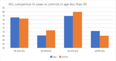
Results: RE Avg RNFL and LE Avg RNFL showed weak correlations with ALSFRS, indicated by Pearson Correlation coefficients of 0.073 and -0.026, respectively. The p-values (0.637 and 0.86) suggested that these correlations were not statistically significant. RE Avg GCL and LE Avg GCL, on the other hand, exhibited moderate positive correlations with ALSFRS scores, with correlation coefficients of 0.337 (RE) and 0.389 (LE). These correlations were statistically significant, as indicated by p-values of 0.021 and 0.006, respectively, suggesting a substantial association between GCL thickness and ALS functional outcomes.
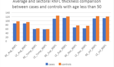
Discussion: All patients in our study were clinically diagnosed cases of ALS, as per the El Escorial criteria. Age group-wise analysis showed statistically significant thinning overall as well as quadrant-wise RNFL parameters in patients less than 50 years compared to age-matched controls, indicating that the pathological process occurring in larger motor neurons in ALS might also be happening in smaller sensory neurons of the retina, causing thinning, which was not due to age-related process. Although GCIPL thinning was occurring in our cases, though statistically not significant compared to control, the significant positive correlation observed between GCIPL and ALS functional outcome and between RNFL and GCIPL measurements highlighted the fact that though the axonal degeneration in retinal neurons might not be translating to the same extent in ganglion cells in ALS, the subtle thinning of GCIPL correlated strongly with functional disability in patients with ALS, implying better functional scores with higher values of GCIPL parameters.
Conclusion: In summary, GCL measurements in both eyes showed a notable relationship with ALSFRS, whereas RNFL did not appear to correlate significantly.

 求助内容:
求助内容: 应助结果提醒方式:


