Vipin Rana, Vikas Sharma, Kanwaljeet Singh, Amit Nandan Tripathi, Ranjit Goenka, Ashish Markan
{"title":"超越通常的怀疑:单侧视盘水肿是Vogt-Koyanagi-Harada综合征的罕见初始体征。","authors":"Vipin Rana, Vikas Sharma, Kanwaljeet Singh, Amit Nandan Tripathi, Ranjit Goenka, Ashish Markan","doi":"10.22336/rjo.2025.44","DOIUrl":null,"url":null,"abstract":"<p><strong>Objective: </strong>To report a case of unilateral optic disc edema as a rare initial presentation of Vogt-Koyanagi-Harada (VKH) syndrome and emphasize the importance of early diagnosis using advanced imaging and cerebrospinal fluid analysis.</p><p><strong>Case presentation: </strong>We present the case of a 23-year-old male who initially presented with unilateral optic disc edema, retro-orbital pain, and headache, progressing to bilateral involvement with serous retinal detachments. Advanced imaging, including fundus fluorescein angiography (FFA), Indocyanine green angiography (ICG), and Enhanced depth imaging-Optical coherence tomography (EDI-OCT), revealed hallmark findings of VKH, such as choroidal granulomas and increased choroidal thickness. Cerebrospinal fluid analysis confirmed pleocytosis and melanin-laden macrophages, which helped to establish the diagnosis. The patient was treated with high-dose intravenous corticosteroids and azathioprine, with significant improvement.</p><p><strong>Discussion: </strong>VKH progresses through prodromal, acute uveitic, chronic, and recurrent phases. Although typically presenting with panuveitis, isolated optic disc edema as an initial sign is rare. Early diagnosis requires multimodal imaging and cerebrospinal fluid analysis to differentiate VKH from other inflammatory and infectious aetiologies.</p><p><strong>Conclusion: </strong>This case highlights the importance of considering VKH in patients presenting with disc edema, whether unilateral or bilateral, particularly when accompanied by vitreous cells. Advanced ocular imaging and thorough systemic evaluation are critical for early diagnosis. Prompt treatment can prevent progression to chronic disease and irreversible vision loss.</p>","PeriodicalId":94355,"journal":{"name":"Romanian journal of ophthalmology","volume":"69 2","pages":"275-279"},"PeriodicalIF":0.0000,"publicationDate":"2025-04-01","publicationTypes":"Journal Article","fieldsOfStudy":null,"isOpenAccess":false,"openAccessPdf":"https://www.ncbi.nlm.nih.gov/pmc/articles/PMC12277989/pdf/","citationCount":"0","resultStr":"{\"title\":\"Beyond the Usual Suspects: Unilateral Optic Disc Edema as a Rare Initial Sign of Vogt-Koyanagi-Harada Syndrome.\",\"authors\":\"Vipin Rana, Vikas Sharma, Kanwaljeet Singh, Amit Nandan Tripathi, Ranjit Goenka, Ashish Markan\",\"doi\":\"10.22336/rjo.2025.44\",\"DOIUrl\":null,\"url\":null,\"abstract\":\"<p><strong>Objective: </strong>To report a case of unilateral optic disc edema as a rare initial presentation of Vogt-Koyanagi-Harada (VKH) syndrome and emphasize the importance of early diagnosis using advanced imaging and cerebrospinal fluid analysis.</p><p><strong>Case presentation: </strong>We present the case of a 23-year-old male who initially presented with unilateral optic disc edema, retro-orbital pain, and headache, progressing to bilateral involvement with serous retinal detachments. Advanced imaging, including fundus fluorescein angiography (FFA), Indocyanine green angiography (ICG), and Enhanced depth imaging-Optical coherence tomography (EDI-OCT), revealed hallmark findings of VKH, such as choroidal granulomas and increased choroidal thickness. Cerebrospinal fluid analysis confirmed pleocytosis and melanin-laden macrophages, which helped to establish the diagnosis. The patient was treated with high-dose intravenous corticosteroids and azathioprine, with significant improvement.</p><p><strong>Discussion: </strong>VKH progresses through prodromal, acute uveitic, chronic, and recurrent phases. Although typically presenting with panuveitis, isolated optic disc edema as an initial sign is rare. Early diagnosis requires multimodal imaging and cerebrospinal fluid analysis to differentiate VKH from other inflammatory and infectious aetiologies.</p><p><strong>Conclusion: </strong>This case highlights the importance of considering VKH in patients presenting with disc edema, whether unilateral or bilateral, particularly when accompanied by vitreous cells. Advanced ocular imaging and thorough systemic evaluation are critical for early diagnosis. Prompt treatment can prevent progression to chronic disease and irreversible vision loss.</p>\",\"PeriodicalId\":94355,\"journal\":{\"name\":\"Romanian journal of ophthalmology\",\"volume\":\"69 2\",\"pages\":\"275-279\"},\"PeriodicalIF\":0.0000,\"publicationDate\":\"2025-04-01\",\"publicationTypes\":\"Journal Article\",\"fieldsOfStudy\":null,\"isOpenAccess\":false,\"openAccessPdf\":\"https://www.ncbi.nlm.nih.gov/pmc/articles/PMC12277989/pdf/\",\"citationCount\":\"0\",\"resultStr\":null,\"platform\":\"Semanticscholar\",\"paperid\":null,\"PeriodicalName\":\"Romanian journal of ophthalmology\",\"FirstCategoryId\":\"1085\",\"ListUrlMain\":\"https://doi.org/10.22336/rjo.2025.44\",\"RegionNum\":0,\"RegionCategory\":null,\"ArticlePicture\":[],\"TitleCN\":null,\"AbstractTextCN\":null,\"PMCID\":null,\"EPubDate\":\"\",\"PubModel\":\"\",\"JCR\":\"\",\"JCRName\":\"\",\"Score\":null,\"Total\":0}","platform":"Semanticscholar","paperid":null,"PeriodicalName":"Romanian journal of ophthalmology","FirstCategoryId":"1085","ListUrlMain":"https://doi.org/10.22336/rjo.2025.44","RegionNum":0,"RegionCategory":null,"ArticlePicture":[],"TitleCN":null,"AbstractTextCN":null,"PMCID":null,"EPubDate":"","PubModel":"","JCR":"","JCRName":"","Score":null,"Total":0}
Beyond the Usual Suspects: Unilateral Optic Disc Edema as a Rare Initial Sign of Vogt-Koyanagi-Harada Syndrome.
Objective: To report a case of unilateral optic disc edema as a rare initial presentation of Vogt-Koyanagi-Harada (VKH) syndrome and emphasize the importance of early diagnosis using advanced imaging and cerebrospinal fluid analysis.
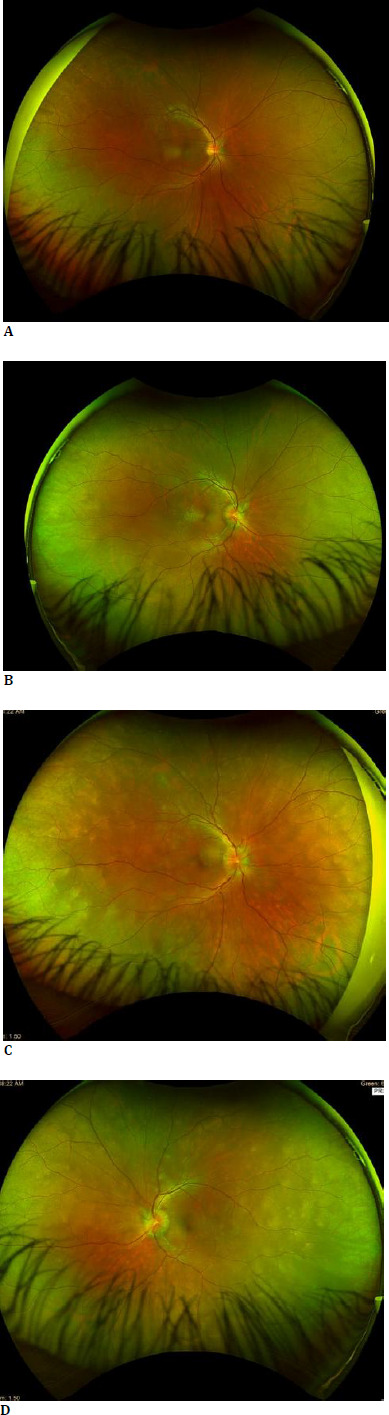
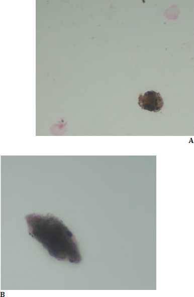
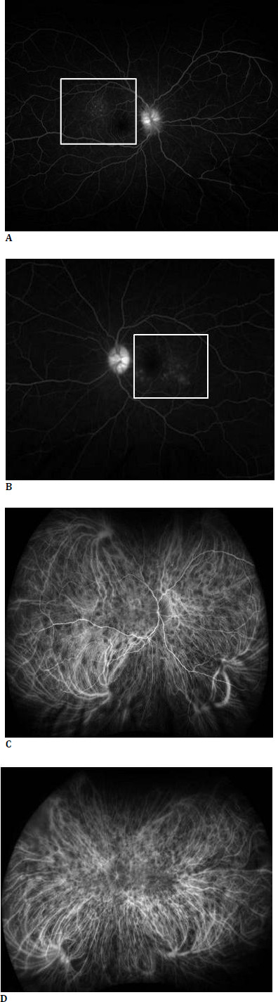
Case presentation: We present the case of a 23-year-old male who initially presented with unilateral optic disc edema, retro-orbital pain, and headache, progressing to bilateral involvement with serous retinal detachments. Advanced imaging, including fundus fluorescein angiography (FFA), Indocyanine green angiography (ICG), and Enhanced depth imaging-Optical coherence tomography (EDI-OCT), revealed hallmark findings of VKH, such as choroidal granulomas and increased choroidal thickness. Cerebrospinal fluid analysis confirmed pleocytosis and melanin-laden macrophages, which helped to establish the diagnosis. The patient was treated with high-dose intravenous corticosteroids and azathioprine, with significant improvement.
Discussion: VKH progresses through prodromal, acute uveitic, chronic, and recurrent phases. Although typically presenting with panuveitis, isolated optic disc edema as an initial sign is rare. Early diagnosis requires multimodal imaging and cerebrospinal fluid analysis to differentiate VKH from other inflammatory and infectious aetiologies.
Conclusion: This case highlights the importance of considering VKH in patients presenting with disc edema, whether unilateral or bilateral, particularly when accompanied by vitreous cells. Advanced ocular imaging and thorough systemic evaluation are critical for early diagnosis. Prompt treatment can prevent progression to chronic disease and irreversible vision loss.

 求助内容:
求助内容: 应助结果提醒方式:


