Baikai Ma, Hongshuo Li, Yi Wang, Wenlong Li, Lei Mou, Yilin Liu, Rongjun Liu, Yalin Zheng, Xinyu Liu, Yitian Zhao, Hong Qi
{"title":"免疫细胞对糖尿病相关性干眼症角膜神经形态学分析的影响及其临床意义。","authors":"Baikai Ma, Hongshuo Li, Yi Wang, Wenlong Li, Lei Mou, Yilin Liu, Rongjun Liu, Yalin Zheng, Xinyu Liu, Yitian Zhao, Hong Qi","doi":"10.1167/tvst.14.7.16","DOIUrl":null,"url":null,"abstract":"<p><strong>Purpose: </strong>Corneal nerve morphology and immune cells are critical biomarkers in the ocular surface. This study aimed to investigate the influence of immune cells on corneal nerve morphology and the clinical significance in diabetes-related dry eye disease (DED).</p><p><strong>Methods: </strong>In the first part, 1075 in vivo confocal microscopy images containing dendritic cells or round cells were included as system validation. Key morphological parameters, including corneal nerve fiber density (CNFD), corneal nerve branch density (CNBD), tortuosity, and box-count fractal dimension (Boxdim), were measured before and after immune cells were excluded. In the second part, a pilot cross-sectional study was conducted involving control (26 eyes), DED without diabetes mellitus (DM) (34 eyes), and DED with DM (17 eyes) groups. The impact of immune cell exclusion on nerve metrics was assessed and correlated with clinical parameters, such as the fluorescein tear breakup time (TBUT), Ocular Surface Disease Index (OSDI), and corneal fluorescein staining (CFS) scores.</p><p><strong>Results: </strong>Exclusion of immune cells resulted in significant reductions in CNFD, CNBD, Boxdim, and tortuosity. Compared to the control group, both DED without DM and DED with DM groups showed substantial reductions in CNFD, CNBD, and Boxdim, along with a significant increase in tortuosity. Moreover, the exclusion of immune cells enhanced the correlations between nerve metrics and fluorescein TBUT.</p><p><strong>Conclusions: </strong>Immune cells contribute to significant biases in the assessments of corneal nerve morphology, primarily false-negative results, in diabetes-related DED. Their exclusion improves the accuracy of nerve measurements, which may enhance the clinical evaluation of corneal nerve morphology.</p><p><strong>Translational relevance: </strong>Advanced segmentation techniques addressing immune cell interference could improve diagnostic precision and inform treatment strategies for DED subtypes.</p>","PeriodicalId":23322,"journal":{"name":"Translational Vision Science & Technology","volume":"14 7","pages":"16"},"PeriodicalIF":2.6000,"publicationDate":"2025-07-01","publicationTypes":"Journal Article","fieldsOfStudy":null,"isOpenAccess":false,"openAccessPdf":"https://www.ncbi.nlm.nih.gov/pmc/articles/PMC12302046/pdf/","citationCount":"0","resultStr":"{\"title\":\"Influence of Immune Cells on Corneal Nerve Morphological Analysis and Clinical Relevance in Diabetes-Related Dry Eye.\",\"authors\":\"Baikai Ma, Hongshuo Li, Yi Wang, Wenlong Li, Lei Mou, Yilin Liu, Rongjun Liu, Yalin Zheng, Xinyu Liu, Yitian Zhao, Hong Qi\",\"doi\":\"10.1167/tvst.14.7.16\",\"DOIUrl\":null,\"url\":null,\"abstract\":\"<p><strong>Purpose: </strong>Corneal nerve morphology and immune cells are critical biomarkers in the ocular surface. This study aimed to investigate the influence of immune cells on corneal nerve morphology and the clinical significance in diabetes-related dry eye disease (DED).</p><p><strong>Methods: </strong>In the first part, 1075 in vivo confocal microscopy images containing dendritic cells or round cells were included as system validation. Key morphological parameters, including corneal nerve fiber density (CNFD), corneal nerve branch density (CNBD), tortuosity, and box-count fractal dimension (Boxdim), were measured before and after immune cells were excluded. In the second part, a pilot cross-sectional study was conducted involving control (26 eyes), DED without diabetes mellitus (DM) (34 eyes), and DED with DM (17 eyes) groups. The impact of immune cell exclusion on nerve metrics was assessed and correlated with clinical parameters, such as the fluorescein tear breakup time (TBUT), Ocular Surface Disease Index (OSDI), and corneal fluorescein staining (CFS) scores.</p><p><strong>Results: </strong>Exclusion of immune cells resulted in significant reductions in CNFD, CNBD, Boxdim, and tortuosity. Compared to the control group, both DED without DM and DED with DM groups showed substantial reductions in CNFD, CNBD, and Boxdim, along with a significant increase in tortuosity. Moreover, the exclusion of immune cells enhanced the correlations between nerve metrics and fluorescein TBUT.</p><p><strong>Conclusions: </strong>Immune cells contribute to significant biases in the assessments of corneal nerve morphology, primarily false-negative results, in diabetes-related DED. Their exclusion improves the accuracy of nerve measurements, which may enhance the clinical evaluation of corneal nerve morphology.</p><p><strong>Translational relevance: </strong>Advanced segmentation techniques addressing immune cell interference could improve diagnostic precision and inform treatment strategies for DED subtypes.</p>\",\"PeriodicalId\":23322,\"journal\":{\"name\":\"Translational Vision Science & Technology\",\"volume\":\"14 7\",\"pages\":\"16\"},\"PeriodicalIF\":2.6000,\"publicationDate\":\"2025-07-01\",\"publicationTypes\":\"Journal Article\",\"fieldsOfStudy\":null,\"isOpenAccess\":false,\"openAccessPdf\":\"https://www.ncbi.nlm.nih.gov/pmc/articles/PMC12302046/pdf/\",\"citationCount\":\"0\",\"resultStr\":null,\"platform\":\"Semanticscholar\",\"paperid\":null,\"PeriodicalName\":\"Translational Vision Science & Technology\",\"FirstCategoryId\":\"3\",\"ListUrlMain\":\"https://doi.org/10.1167/tvst.14.7.16\",\"RegionNum\":3,\"RegionCategory\":\"医学\",\"ArticlePicture\":[],\"TitleCN\":null,\"AbstractTextCN\":null,\"PMCID\":null,\"EPubDate\":\"\",\"PubModel\":\"\",\"JCR\":\"Q2\",\"JCRName\":\"OPHTHALMOLOGY\",\"Score\":null,\"Total\":0}","platform":"Semanticscholar","paperid":null,"PeriodicalName":"Translational Vision Science & Technology","FirstCategoryId":"3","ListUrlMain":"https://doi.org/10.1167/tvst.14.7.16","RegionNum":3,"RegionCategory":"医学","ArticlePicture":[],"TitleCN":null,"AbstractTextCN":null,"PMCID":null,"EPubDate":"","PubModel":"","JCR":"Q2","JCRName":"OPHTHALMOLOGY","Score":null,"Total":0}
Influence of Immune Cells on Corneal Nerve Morphological Analysis and Clinical Relevance in Diabetes-Related Dry Eye.
Purpose: Corneal nerve morphology and immune cells are critical biomarkers in the ocular surface. This study aimed to investigate the influence of immune cells on corneal nerve morphology and the clinical significance in diabetes-related dry eye disease (DED).
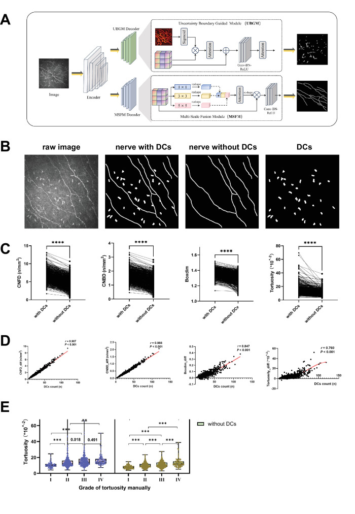
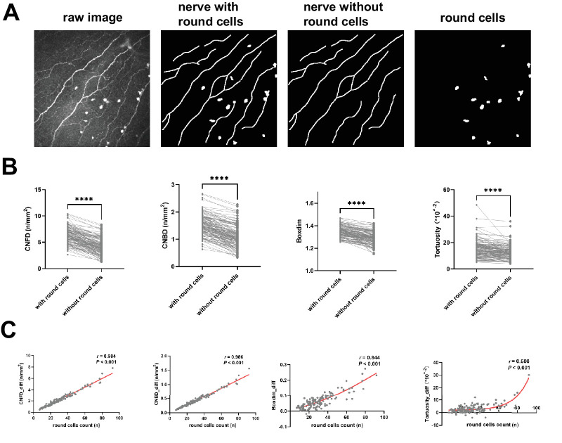
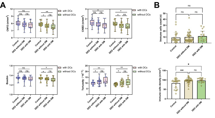
Methods: In the first part, 1075 in vivo confocal microscopy images containing dendritic cells or round cells were included as system validation. Key morphological parameters, including corneal nerve fiber density (CNFD), corneal nerve branch density (CNBD), tortuosity, and box-count fractal dimension (Boxdim), were measured before and after immune cells were excluded. In the second part, a pilot cross-sectional study was conducted involving control (26 eyes), DED without diabetes mellitus (DM) (34 eyes), and DED with DM (17 eyes) groups. The impact of immune cell exclusion on nerve metrics was assessed and correlated with clinical parameters, such as the fluorescein tear breakup time (TBUT), Ocular Surface Disease Index (OSDI), and corneal fluorescein staining (CFS) scores.
Results: Exclusion of immune cells resulted in significant reductions in CNFD, CNBD, Boxdim, and tortuosity. Compared to the control group, both DED without DM and DED with DM groups showed substantial reductions in CNFD, CNBD, and Boxdim, along with a significant increase in tortuosity. Moreover, the exclusion of immune cells enhanced the correlations between nerve metrics and fluorescein TBUT.
Conclusions: Immune cells contribute to significant biases in the assessments of corneal nerve morphology, primarily false-negative results, in diabetes-related DED. Their exclusion improves the accuracy of nerve measurements, which may enhance the clinical evaluation of corneal nerve morphology.
Translational relevance: Advanced segmentation techniques addressing immune cell interference could improve diagnostic precision and inform treatment strategies for DED subtypes.
期刊介绍:
Translational Vision Science & Technology (TVST), an official journal of the Association for Research in Vision and Ophthalmology (ARVO), an international organization whose purpose is to advance research worldwide into understanding the visual system and preventing, treating and curing its disorders, is an online, open access, peer-reviewed journal emphasizing multidisciplinary research that bridges the gap between basic research and clinical care. A highly qualified and diverse group of Associate Editors and Editorial Board Members is led by Editor-in-Chief Marco Zarbin, MD, PhD, FARVO.
The journal covers a broad spectrum of work, including but not limited to:
Applications of stem cell technology for regenerative medicine,
Development of new animal models of human diseases,
Tissue bioengineering,
Chemical engineering to improve virus-based gene delivery,
Nanotechnology for drug delivery,
Design and synthesis of artificial extracellular matrices,
Development of a true microsurgical operating environment,
Refining data analysis algorithms to improve in vivo imaging technology,
Results of Phase 1 clinical trials,
Reverse translational ("bedside to bench") research.
TVST seeks manuscripts from scientists and clinicians with diverse backgrounds ranging from basic chemistry to ophthalmic surgery that will advance or change the way we understand and/or treat vision-threatening diseases. TVST encourages the use of color, multimedia, hyperlinks, program code and other digital enhancements.

 求助内容:
求助内容: 应助结果提醒方式:


