Cassio V Ruas, Bruna M Carlos, Saulo Feitosa, Márcio Vinícius Silva, Pedro Vazquez, Larissa L Pontes, Jayne Silvestre, Sara R M Almeida, Alexandre F Brandão, Gabriela Castellano
{"title":"扩展现实训练中经颅直流电刺激对脑卒中幸存者皮层、神经肌肉和临床恢复的影响。","authors":"Cassio V Ruas, Bruna M Carlos, Saulo Feitosa, Márcio Vinícius Silva, Pedro Vazquez, Larissa L Pontes, Jayne Silvestre, Sara R M Almeida, Alexandre F Brandão, Gabriela Castellano","doi":"10.1155/np/5688648","DOIUrl":null,"url":null,"abstract":"<p><p><b>Background:</b> Rehabilitation methods that include anodal transcranial direct current stimulation (<i>a</i>tDCS) and extended reality (XR) exercises have been used to enhance neural networks and improve functional performance in stroke patients, but the neuromuscular and neurophysiological mechanisms underlying these improvements are not fully understood. The purpose of this study was to examine the effects of <i>a</i>tDCS during XR rehabilitation exercises on cortical, neuromuscular, and clinical outcomes of stroke survivors. <b>Methods:</b> Nineteen chronic stroke survivors were placed into either a transcranial direct current stimulation (tDCS) or a <i>Sham</i> group, without significant (<i>p</i> > 0.73) differences in the baseline levels of disability between groups. The tDCS group received active <i>a</i>tDCS and the <i>Sham</i> group received sham <i>a</i>tDCS applied on the ipsilesional primary motor cortex (M1) while performing a 10-session XR rehabilitation program. Surface electromyography (EMG) activity was recorded from deltoid and rectus femoris of the paretic limb without and with the application of active/sham <i>a</i>tDCS on the M1. Shoulder abduction and hip flexion active maximum joint range of motion (ROM<sub>max</sub>), electroencephalography (EEG)-derived brain symmetry index (BSI) and functional/clinical tests were assessed before and after the rehabilitation program. <b>Results:</b> EMG activity was ~ 31% greater during hip flexion of the paretic limb with the application of active <i>a</i>tDCS than without <i>a</i>tDCS (<i>p</i>=0.04). Paretic hip flexion ROM<sub>max</sub> increased by ~ 26%, BSI decreased by ~ 72% (indicating greater brain symmetry) and timed up and go (TUG) functional test improved by ~ 11% from before to after the rehabilitation program for the tDCS group only (<i>p</i> < 0.05). No other significant differences (<i>p</i> > 0.05) were observed. <b>Conclusion:</b> It seems that the application of active <i>a</i>tDCS targeted the ipsilesional M1 representation of the quadriceps, which potentiated muscle activation in the paretic rectus femoris during XR exercises and resulted in greater motor recovery in hip flexion movements. The EEG-derived BSI results also indicate that <i>a</i>tDCS was effective in reorganizing the ipsilesional hemisphere brain activity after stroke.</p>","PeriodicalId":19122,"journal":{"name":"Neural Plasticity","volume":"2025 ","pages":"5688648"},"PeriodicalIF":3.7000,"publicationDate":"2025-07-15","publicationTypes":"Journal Article","fieldsOfStudy":null,"isOpenAccess":false,"openAccessPdf":"https://www.ncbi.nlm.nih.gov/pmc/articles/PMC12283194/pdf/","citationCount":"0","resultStr":"{\"title\":\"The Effects of Transcranial Direct Current Stimulation During Extended Reality Exercises for Cortical, Neuromuscular, and Clinical Recovery of Stroke Survivors.\",\"authors\":\"Cassio V Ruas, Bruna M Carlos, Saulo Feitosa, Márcio Vinícius Silva, Pedro Vazquez, Larissa L Pontes, Jayne Silvestre, Sara R M Almeida, Alexandre F Brandão, Gabriela Castellano\",\"doi\":\"10.1155/np/5688648\",\"DOIUrl\":null,\"url\":null,\"abstract\":\"<p><p><b>Background:</b> Rehabilitation methods that include anodal transcranial direct current stimulation (<i>a</i>tDCS) and extended reality (XR) exercises have been used to enhance neural networks and improve functional performance in stroke patients, but the neuromuscular and neurophysiological mechanisms underlying these improvements are not fully understood. The purpose of this study was to examine the effects of <i>a</i>tDCS during XR rehabilitation exercises on cortical, neuromuscular, and clinical outcomes of stroke survivors. <b>Methods:</b> Nineteen chronic stroke survivors were placed into either a transcranial direct current stimulation (tDCS) or a <i>Sham</i> group, without significant (<i>p</i> > 0.73) differences in the baseline levels of disability between groups. The tDCS group received active <i>a</i>tDCS and the <i>Sham</i> group received sham <i>a</i>tDCS applied on the ipsilesional primary motor cortex (M1) while performing a 10-session XR rehabilitation program. Surface electromyography (EMG) activity was recorded from deltoid and rectus femoris of the paretic limb without and with the application of active/sham <i>a</i>tDCS on the M1. Shoulder abduction and hip flexion active maximum joint range of motion (ROM<sub>max</sub>), electroencephalography (EEG)-derived brain symmetry index (BSI) and functional/clinical tests were assessed before and after the rehabilitation program. <b>Results:</b> EMG activity was ~ 31% greater during hip flexion of the paretic limb with the application of active <i>a</i>tDCS than without <i>a</i>tDCS (<i>p</i>=0.04). Paretic hip flexion ROM<sub>max</sub> increased by ~ 26%, BSI decreased by ~ 72% (indicating greater brain symmetry) and timed up and go (TUG) functional test improved by ~ 11% from before to after the rehabilitation program for the tDCS group only (<i>p</i> < 0.05). No other significant differences (<i>p</i> > 0.05) were observed. <b>Conclusion:</b> It seems that the application of active <i>a</i>tDCS targeted the ipsilesional M1 representation of the quadriceps, which potentiated muscle activation in the paretic rectus femoris during XR exercises and resulted in greater motor recovery in hip flexion movements. The EEG-derived BSI results also indicate that <i>a</i>tDCS was effective in reorganizing the ipsilesional hemisphere brain activity after stroke.</p>\",\"PeriodicalId\":19122,\"journal\":{\"name\":\"Neural Plasticity\",\"volume\":\"2025 \",\"pages\":\"5688648\"},\"PeriodicalIF\":3.7000,\"publicationDate\":\"2025-07-15\",\"publicationTypes\":\"Journal Article\",\"fieldsOfStudy\":null,\"isOpenAccess\":false,\"openAccessPdf\":\"https://www.ncbi.nlm.nih.gov/pmc/articles/PMC12283194/pdf/\",\"citationCount\":\"0\",\"resultStr\":null,\"platform\":\"Semanticscholar\",\"paperid\":null,\"PeriodicalName\":\"Neural Plasticity\",\"FirstCategoryId\":\"3\",\"ListUrlMain\":\"https://doi.org/10.1155/np/5688648\",\"RegionNum\":4,\"RegionCategory\":\"医学\",\"ArticlePicture\":[],\"TitleCN\":null,\"AbstractTextCN\":null,\"PMCID\":null,\"EPubDate\":\"2025/1/1 0:00:00\",\"PubModel\":\"eCollection\",\"JCR\":\"Q2\",\"JCRName\":\"Medicine\",\"Score\":null,\"Total\":0}","platform":"Semanticscholar","paperid":null,"PeriodicalName":"Neural Plasticity","FirstCategoryId":"3","ListUrlMain":"https://doi.org/10.1155/np/5688648","RegionNum":4,"RegionCategory":"医学","ArticlePicture":[],"TitleCN":null,"AbstractTextCN":null,"PMCID":null,"EPubDate":"2025/1/1 0:00:00","PubModel":"eCollection","JCR":"Q2","JCRName":"Medicine","Score":null,"Total":0}
引用次数: 0
摘要
背景:包括阳极经颅直流电刺激(atDCS)和扩展现实(XR)练习在内的康复方法已被用于增强脑卒中患者的神经网络和改善功能表现,但这些改善背后的神经肌肉和神经生理机制尚不完全清楚。本研究的目的是研究在XR康复训练中atDCS对脑卒中幸存者皮质、神经肌肉和临床结果的影响。方法:19名慢性脑卒中幸存者被分为经颅直流电刺激(tDCS)组和假手术组,两组之间残疾基线水平无显著差异(p > 0.73)。tDCS组接受活性atDCS治疗,Sham组接受假atDCS治疗,同时进行10次XR康复计划。在没有和使用主动/假性dcs的情况下,记录瘫肢体的三角肌和股直肌的表面肌电(EMG)活动。在康复计划前后评估肩部外展和髋关节屈曲活动最大关节活动范围(ROMmax),脑电图(EEG)衍生的脑对称指数(BSI)和功能/临床测试。结果:与未应用atDCS相比,应用活动atDCS的瘫瘫肢体髋关节屈曲时肌电活动增加约31% (p=0.04)。仅tDCS组的瘫瘫性髋屈曲ROMmax比康复前提高了~ 26%,BSI降低了~ 72%(表明脑对称性增强),而时间up and go (TUG)功能测试比康复前提高了~ 11% (p < 0.05)。其他差异无统计学意义(p < 0.05)。结论:活动性atDCS的应用似乎是针对股四头肌的同侧M1表现,在XR运动中增强了麻痹性股直肌的肌肉激活,并导致髋关节屈曲运动中更大的运动恢复。脑电衍生的BSI结果也表明,atDCS对卒中后同侧半球大脑活动的重组是有效的。
The Effects of Transcranial Direct Current Stimulation During Extended Reality Exercises for Cortical, Neuromuscular, and Clinical Recovery of Stroke Survivors.
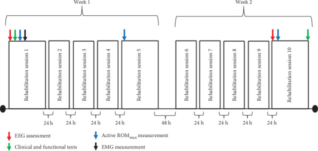
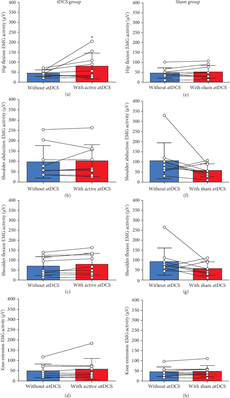
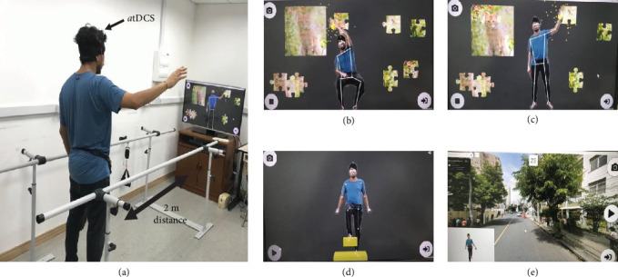
Background: Rehabilitation methods that include anodal transcranial direct current stimulation (atDCS) and extended reality (XR) exercises have been used to enhance neural networks and improve functional performance in stroke patients, but the neuromuscular and neurophysiological mechanisms underlying these improvements are not fully understood. The purpose of this study was to examine the effects of atDCS during XR rehabilitation exercises on cortical, neuromuscular, and clinical outcomes of stroke survivors. Methods: Nineteen chronic stroke survivors were placed into either a transcranial direct current stimulation (tDCS) or a Sham group, without significant (p > 0.73) differences in the baseline levels of disability between groups. The tDCS group received active atDCS and the Sham group received sham atDCS applied on the ipsilesional primary motor cortex (M1) while performing a 10-session XR rehabilitation program. Surface electromyography (EMG) activity was recorded from deltoid and rectus femoris of the paretic limb without and with the application of active/sham atDCS on the M1. Shoulder abduction and hip flexion active maximum joint range of motion (ROMmax), electroencephalography (EEG)-derived brain symmetry index (BSI) and functional/clinical tests were assessed before and after the rehabilitation program. Results: EMG activity was ~ 31% greater during hip flexion of the paretic limb with the application of active atDCS than without atDCS (p=0.04). Paretic hip flexion ROMmax increased by ~ 26%, BSI decreased by ~ 72% (indicating greater brain symmetry) and timed up and go (TUG) functional test improved by ~ 11% from before to after the rehabilitation program for the tDCS group only (p < 0.05). No other significant differences (p > 0.05) were observed. Conclusion: It seems that the application of active atDCS targeted the ipsilesional M1 representation of the quadriceps, which potentiated muscle activation in the paretic rectus femoris during XR exercises and resulted in greater motor recovery in hip flexion movements. The EEG-derived BSI results also indicate that atDCS was effective in reorganizing the ipsilesional hemisphere brain activity after stroke.
期刊介绍:
Neural Plasticity is an international, interdisciplinary journal dedicated to the publication of articles related to all aspects of neural plasticity, with special emphasis on its functional significance as reflected in behavior and in psychopathology. Neural Plasticity publishes research and review articles from the entire range of relevant disciplines, including basic neuroscience, behavioral neuroscience, cognitive neuroscience, biological psychology, and biological psychiatry.
 求助内容:
求助内容: 应助结果提醒方式:


