Duaa Knaj, Ghanem Ahmad, Zuheir Alshehabi, Issa Yusuof Ahmad
{"title":"腓骨转移至肺部的金刚烷瘤:一罕见病例报告。","authors":"Duaa Knaj, Ghanem Ahmad, Zuheir Alshehabi, Issa Yusuof Ahmad","doi":"10.2147/IMCRJ.S535403","DOIUrl":null,"url":null,"abstract":"<p><strong>Background: </strong>Adamantinoma is a low-grade, primary malignant bone tumor commonly found in the tibia with or without fibular involvement.</p><p><strong>Case presentation: </strong>A 21-year-old female patient was admitted to the hospital with a mass on the upper lateral aspect of the left leg. She reported the onset of a small swelling in the left leg eight months prior to admission. It has gradually increased in size to involve the upper leg and part of the knee. The patient had limited range of motion in the knee joint and difficulty leaning on the affected limb. Radiographs showed a cortical-destroying osteolytic lesion at the head of the fibula extending into the surrounding soft tissue. Ultrasound imaging showed a mixed echo formation with indistinct borders and multiple chambers separated by trabeculae demonstrating blood flow. MRI revealed the absence of the fibular head as evidenced by cortical destruction and extension into adjacent soft tissue. CT scan showed a 12×13 cm mass in the left popliteal region extending to the knee margin. Enlarged lymph nodes were noted in the left groin. Multiple small metastases were observed in the lungs. Histologically, atypical epithelial cells and stromal spindle cells were observed. CK and CK19 staining was positive. The patient was treated with Sunitinib 50 mg orally, once daily for 4 weeks, followed by 2 weeks off for 2 cycles. During treatment (after 2 cycles) the tumor showed progressive growth, resulting in decreased function of the affected limb. She was referred to the surgical department where the appropriate surgical amputation was performed. After surgery, the patient's general condition improved. A further CT scan revealed a slight progression of metastatic lesions in the lungs, which prompted the decision to administer chemotherapy, but she refused to continue the treatment and did not receive chemotherapy. Two months later, she presented to the hospital in poor general condition. CT scan revealed extensive and progressive lesions in both lungs, leading to chest pain, hemoptysis, and hypoxia. Despite supportive care, the patient eventually died.</p><p><strong>Conclusion: </strong>This case report presents a rare case of metastatic adamantinoma that did not respond to treatment with Sunitinib.</p>","PeriodicalId":14337,"journal":{"name":"International Medical Case Reports Journal","volume":"18 ","pages":"909-914"},"PeriodicalIF":0.7000,"publicationDate":"2025-07-18","publicationTypes":"Journal Article","fieldsOfStudy":null,"isOpenAccess":false,"openAccessPdf":"https://www.ncbi.nlm.nih.gov/pmc/articles/PMC12282602/pdf/","citationCount":"0","resultStr":"{\"title\":\"Metastatic Adamantinoma of Fibula to the Lungs: A Rare Case Report.\",\"authors\":\"Duaa Knaj, Ghanem Ahmad, Zuheir Alshehabi, Issa Yusuof Ahmad\",\"doi\":\"10.2147/IMCRJ.S535403\",\"DOIUrl\":null,\"url\":null,\"abstract\":\"<p><strong>Background: </strong>Adamantinoma is a low-grade, primary malignant bone tumor commonly found in the tibia with or without fibular involvement.</p><p><strong>Case presentation: </strong>A 21-year-old female patient was admitted to the hospital with a mass on the upper lateral aspect of the left leg. She reported the onset of a small swelling in the left leg eight months prior to admission. It has gradually increased in size to involve the upper leg and part of the knee. The patient had limited range of motion in the knee joint and difficulty leaning on the affected limb. Radiographs showed a cortical-destroying osteolytic lesion at the head of the fibula extending into the surrounding soft tissue. Ultrasound imaging showed a mixed echo formation with indistinct borders and multiple chambers separated by trabeculae demonstrating blood flow. MRI revealed the absence of the fibular head as evidenced by cortical destruction and extension into adjacent soft tissue. CT scan showed a 12×13 cm mass in the left popliteal region extending to the knee margin. Enlarged lymph nodes were noted in the left groin. Multiple small metastases were observed in the lungs. Histologically, atypical epithelial cells and stromal spindle cells were observed. CK and CK19 staining was positive. The patient was treated with Sunitinib 50 mg orally, once daily for 4 weeks, followed by 2 weeks off for 2 cycles. During treatment (after 2 cycles) the tumor showed progressive growth, resulting in decreased function of the affected limb. She was referred to the surgical department where the appropriate surgical amputation was performed. After surgery, the patient's general condition improved. A further CT scan revealed a slight progression of metastatic lesions in the lungs, which prompted the decision to administer chemotherapy, but she refused to continue the treatment and did not receive chemotherapy. Two months later, she presented to the hospital in poor general condition. CT scan revealed extensive and progressive lesions in both lungs, leading to chest pain, hemoptysis, and hypoxia. Despite supportive care, the patient eventually died.</p><p><strong>Conclusion: </strong>This case report presents a rare case of metastatic adamantinoma that did not respond to treatment with Sunitinib.</p>\",\"PeriodicalId\":14337,\"journal\":{\"name\":\"International Medical Case Reports Journal\",\"volume\":\"18 \",\"pages\":\"909-914\"},\"PeriodicalIF\":0.7000,\"publicationDate\":\"2025-07-18\",\"publicationTypes\":\"Journal Article\",\"fieldsOfStudy\":null,\"isOpenAccess\":false,\"openAccessPdf\":\"https://www.ncbi.nlm.nih.gov/pmc/articles/PMC12282602/pdf/\",\"citationCount\":\"0\",\"resultStr\":null,\"platform\":\"Semanticscholar\",\"paperid\":null,\"PeriodicalName\":\"International Medical Case Reports Journal\",\"FirstCategoryId\":\"1085\",\"ListUrlMain\":\"https://doi.org/10.2147/IMCRJ.S535403\",\"RegionNum\":0,\"RegionCategory\":null,\"ArticlePicture\":[],\"TitleCN\":null,\"AbstractTextCN\":null,\"PMCID\":null,\"EPubDate\":\"2025/1/1 0:00:00\",\"PubModel\":\"eCollection\",\"JCR\":\"Q3\",\"JCRName\":\"MEDICINE, GENERAL & INTERNAL\",\"Score\":null,\"Total\":0}","platform":"Semanticscholar","paperid":null,"PeriodicalName":"International Medical Case Reports Journal","FirstCategoryId":"1085","ListUrlMain":"https://doi.org/10.2147/IMCRJ.S535403","RegionNum":0,"RegionCategory":null,"ArticlePicture":[],"TitleCN":null,"AbstractTextCN":null,"PMCID":null,"EPubDate":"2025/1/1 0:00:00","PubModel":"eCollection","JCR":"Q3","JCRName":"MEDICINE, GENERAL & INTERNAL","Score":null,"Total":0}
Metastatic Adamantinoma of Fibula to the Lungs: A Rare Case Report.
Background: Adamantinoma is a low-grade, primary malignant bone tumor commonly found in the tibia with or without fibular involvement.
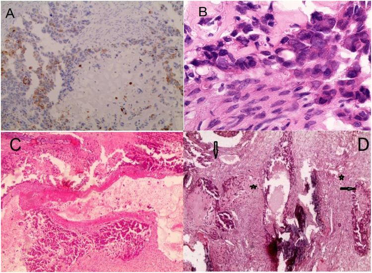
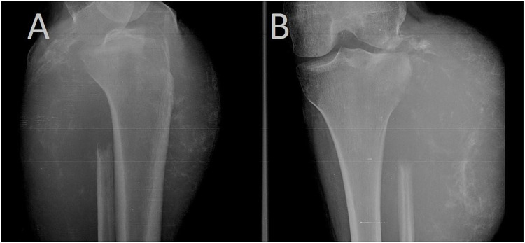
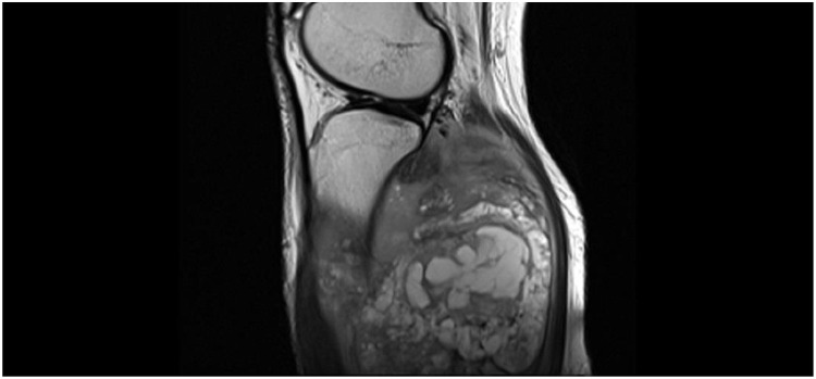
Case presentation: A 21-year-old female patient was admitted to the hospital with a mass on the upper lateral aspect of the left leg. She reported the onset of a small swelling in the left leg eight months prior to admission. It has gradually increased in size to involve the upper leg and part of the knee. The patient had limited range of motion in the knee joint and difficulty leaning on the affected limb. Radiographs showed a cortical-destroying osteolytic lesion at the head of the fibula extending into the surrounding soft tissue. Ultrasound imaging showed a mixed echo formation with indistinct borders and multiple chambers separated by trabeculae demonstrating blood flow. MRI revealed the absence of the fibular head as evidenced by cortical destruction and extension into adjacent soft tissue. CT scan showed a 12×13 cm mass in the left popliteal region extending to the knee margin. Enlarged lymph nodes were noted in the left groin. Multiple small metastases were observed in the lungs. Histologically, atypical epithelial cells and stromal spindle cells were observed. CK and CK19 staining was positive. The patient was treated with Sunitinib 50 mg orally, once daily for 4 weeks, followed by 2 weeks off for 2 cycles. During treatment (after 2 cycles) the tumor showed progressive growth, resulting in decreased function of the affected limb. She was referred to the surgical department where the appropriate surgical amputation was performed. After surgery, the patient's general condition improved. A further CT scan revealed a slight progression of metastatic lesions in the lungs, which prompted the decision to administer chemotherapy, but she refused to continue the treatment and did not receive chemotherapy. Two months later, she presented to the hospital in poor general condition. CT scan revealed extensive and progressive lesions in both lungs, leading to chest pain, hemoptysis, and hypoxia. Despite supportive care, the patient eventually died.
Conclusion: This case report presents a rare case of metastatic adamantinoma that did not respond to treatment with Sunitinib.
期刊介绍:
International Medical Case Reports Journal is an international, peer-reviewed, open access, online journal publishing original case reports from all medical specialties. Submissions should not normally exceed 3,000 words or 4 published pages including figures, diagrams and references. As of 1st April 2019, the International Medical Case Reports Journal will no longer consider meta-analyses for publication.

 求助内容:
求助内容: 应助结果提醒方式:


