Simon Schading-Sassenhausen, Anna Lebret, Kadir Şimşek, Pauline Gut, Sabrina Imhof, Björn Zörner, Roland Kreis, Patrick Freund, Maryam Seif
{"title":"脊髓损伤后运动系统的代谢和结构改变:体内1H-MR光谱研究","authors":"Simon Schading-Sassenhausen, Anna Lebret, Kadir Şimşek, Pauline Gut, Sabrina Imhof, Björn Zörner, Roland Kreis, Patrick Freund, Maryam Seif","doi":"10.1002/jnr.70071","DOIUrl":null,"url":null,"abstract":"<p>Spinal cord injury (SCI) disrupts spinal tracts and neuronal pathways, including those in the primary motor cortex (M1) and the lumbar cord enlargement (LCE) involved in motor control. This study sought to determine whether metabolite concentrations deviate between SCI and healthy controls (HC) in M1 and LCE using proton magnetic resonance spectroscopy (<sup>1</sup>H-MRS) and structural MRI, and if these correlate with clinical impairment. Sixteen chronic SCI (mean age: 54.7 ± 14.8y) and 19 HCs (mean age: 53.2 ± 18.8y) underwent <sup>1</sup>H-MRS to quantify metabolites along with T<sub>1</sub>- and T<sub>2</sub>*-weighted MRI to assess tissue structural changes. Associations between metabolic and structural changes and clinical impairment were also assessed. Patients showed significant atrophy in both white matter of the LCE (HC: 37.7 ± 4.7 mm<sup>2</sup>, SCI: 33.9 ± 3.7 mm<sup>2</sup>, Δ = −10.1%, <i>p</i> = 0.015) and gray matter (HC: 20.9 ± 2.1 mm<sup>2</sup>, SCI: 19.4 ± 1.5 mm<sup>2</sup>, Δ = −7.2%, <i>p</i> = 0.022). Total N-acetylaspartate (tNAA) with respect to total creatine (tCr) was reduced in M1 of SCI (HC: 1.94 ± 0.21, SCI: 1.77 ± 0.14, ∆ = −8.8%, <i>p</i> = 0.006) and in the LCE (HC: 2.48 ± 0.76, SCI: 1.81 ± 0.80, ∆ = −27.0%, <i>p</i> = 0.02). In conclusion, reduced tNAA/tCr in both the atrophied LCE and M1 suggests widespread neuronal changes including cell atrophy and/or cell loss after injury. These findings provide in vivo evidence for retrograde and trans-synaptic neurodegeneration, which may underline the atrophy observed in the motor system in SCI. Ultimately, this highlights the potential for metabolic and structural biomarkers to improve the monitoring of subtle neurodegeneration following SCI and to enhance future regenerative treatment strategies.</p>","PeriodicalId":16490,"journal":{"name":"Journal of Neuroscience Research","volume":"103 7","pages":""},"PeriodicalIF":3.4000,"publicationDate":"2025-07-24","publicationTypes":"Journal Article","fieldsOfStudy":null,"isOpenAccess":false,"openAccessPdf":"https://onlinelibrary.wiley.com/doi/epdf/10.1002/jnr.70071","citationCount":"0","resultStr":"{\"title\":\"Metabolic and Structural Alterations in the Motor System Following Spinal Cord Injury: An In-Vivo 1H-MR Spectroscopy Investigation\",\"authors\":\"Simon Schading-Sassenhausen, Anna Lebret, Kadir Şimşek, Pauline Gut, Sabrina Imhof, Björn Zörner, Roland Kreis, Patrick Freund, Maryam Seif\",\"doi\":\"10.1002/jnr.70071\",\"DOIUrl\":null,\"url\":null,\"abstract\":\"<p>Spinal cord injury (SCI) disrupts spinal tracts and neuronal pathways, including those in the primary motor cortex (M1) and the lumbar cord enlargement (LCE) involved in motor control. This study sought to determine whether metabolite concentrations deviate between SCI and healthy controls (HC) in M1 and LCE using proton magnetic resonance spectroscopy (<sup>1</sup>H-MRS) and structural MRI, and if these correlate with clinical impairment. Sixteen chronic SCI (mean age: 54.7 ± 14.8y) and 19 HCs (mean age: 53.2 ± 18.8y) underwent <sup>1</sup>H-MRS to quantify metabolites along with T<sub>1</sub>- and T<sub>2</sub>*-weighted MRI to assess tissue structural changes. Associations between metabolic and structural changes and clinical impairment were also assessed. Patients showed significant atrophy in both white matter of the LCE (HC: 37.7 ± 4.7 mm<sup>2</sup>, SCI: 33.9 ± 3.7 mm<sup>2</sup>, Δ = −10.1%, <i>p</i> = 0.015) and gray matter (HC: 20.9 ± 2.1 mm<sup>2</sup>, SCI: 19.4 ± 1.5 mm<sup>2</sup>, Δ = −7.2%, <i>p</i> = 0.022). Total N-acetylaspartate (tNAA) with respect to total creatine (tCr) was reduced in M1 of SCI (HC: 1.94 ± 0.21, SCI: 1.77 ± 0.14, ∆ = −8.8%, <i>p</i> = 0.006) and in the LCE (HC: 2.48 ± 0.76, SCI: 1.81 ± 0.80, ∆ = −27.0%, <i>p</i> = 0.02). In conclusion, reduced tNAA/tCr in both the atrophied LCE and M1 suggests widespread neuronal changes including cell atrophy and/or cell loss after injury. These findings provide in vivo evidence for retrograde and trans-synaptic neurodegeneration, which may underline the atrophy observed in the motor system in SCI. Ultimately, this highlights the potential for metabolic and structural biomarkers to improve the monitoring of subtle neurodegeneration following SCI and to enhance future regenerative treatment strategies.</p>\",\"PeriodicalId\":16490,\"journal\":{\"name\":\"Journal of Neuroscience Research\",\"volume\":\"103 7\",\"pages\":\"\"},\"PeriodicalIF\":3.4000,\"publicationDate\":\"2025-07-24\",\"publicationTypes\":\"Journal Article\",\"fieldsOfStudy\":null,\"isOpenAccess\":false,\"openAccessPdf\":\"https://onlinelibrary.wiley.com/doi/epdf/10.1002/jnr.70071\",\"citationCount\":\"0\",\"resultStr\":null,\"platform\":\"Semanticscholar\",\"paperid\":null,\"PeriodicalName\":\"Journal of Neuroscience Research\",\"FirstCategoryId\":\"3\",\"ListUrlMain\":\"https://onlinelibrary.wiley.com/doi/10.1002/jnr.70071\",\"RegionNum\":3,\"RegionCategory\":\"医学\",\"ArticlePicture\":[],\"TitleCN\":null,\"AbstractTextCN\":null,\"PMCID\":null,\"EPubDate\":\"\",\"PubModel\":\"\",\"JCR\":\"Q2\",\"JCRName\":\"NEUROSCIENCES\",\"Score\":null,\"Total\":0}","platform":"Semanticscholar","paperid":null,"PeriodicalName":"Journal of Neuroscience Research","FirstCategoryId":"3","ListUrlMain":"https://onlinelibrary.wiley.com/doi/10.1002/jnr.70071","RegionNum":3,"RegionCategory":"医学","ArticlePicture":[],"TitleCN":null,"AbstractTextCN":null,"PMCID":null,"EPubDate":"","PubModel":"","JCR":"Q2","JCRName":"NEUROSCIENCES","Score":null,"Total":0}
Metabolic and Structural Alterations in the Motor System Following Spinal Cord Injury: An In-Vivo 1H-MR Spectroscopy Investigation
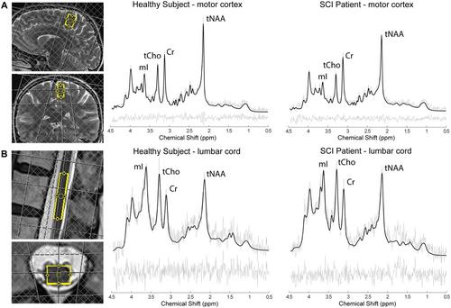
Spinal cord injury (SCI) disrupts spinal tracts and neuronal pathways, including those in the primary motor cortex (M1) and the lumbar cord enlargement (LCE) involved in motor control. This study sought to determine whether metabolite concentrations deviate between SCI and healthy controls (HC) in M1 and LCE using proton magnetic resonance spectroscopy (1H-MRS) and structural MRI, and if these correlate with clinical impairment. Sixteen chronic SCI (mean age: 54.7 ± 14.8y) and 19 HCs (mean age: 53.2 ± 18.8y) underwent 1H-MRS to quantify metabolites along with T1- and T2*-weighted MRI to assess tissue structural changes. Associations between metabolic and structural changes and clinical impairment were also assessed. Patients showed significant atrophy in both white matter of the LCE (HC: 37.7 ± 4.7 mm2, SCI: 33.9 ± 3.7 mm2, Δ = −10.1%, p = 0.015) and gray matter (HC: 20.9 ± 2.1 mm2, SCI: 19.4 ± 1.5 mm2, Δ = −7.2%, p = 0.022). Total N-acetylaspartate (tNAA) with respect to total creatine (tCr) was reduced in M1 of SCI (HC: 1.94 ± 0.21, SCI: 1.77 ± 0.14, ∆ = −8.8%, p = 0.006) and in the LCE (HC: 2.48 ± 0.76, SCI: 1.81 ± 0.80, ∆ = −27.0%, p = 0.02). In conclusion, reduced tNAA/tCr in both the atrophied LCE and M1 suggests widespread neuronal changes including cell atrophy and/or cell loss after injury. These findings provide in vivo evidence for retrograde and trans-synaptic neurodegeneration, which may underline the atrophy observed in the motor system in SCI. Ultimately, this highlights the potential for metabolic and structural biomarkers to improve the monitoring of subtle neurodegeneration following SCI and to enhance future regenerative treatment strategies.
期刊介绍:
The Journal of Neuroscience Research (JNR) publishes novel research results that will advance our understanding of the development, function and pathophysiology of the nervous system, using molecular, cellular, systems, and translational approaches. JNR covers both basic research and clinical aspects of neurology, neuropathology, psychiatry or psychology.
The journal focuses on uncovering the intricacies of brain structure and function. Research published in JNR covers all species from invertebrates to humans, and the reports inform the readers about the function and organization of the nervous system, with emphasis on how disease modifies the function and organization.
 求助内容:
求助内容: 应助结果提醒方式:


