丁香假单胞菌细胞壁生物聚合物动力学的拉曼成像化学计量学分析。猕猴桃茎被猕猴桃酸菌感染

IF 1.3 4区 化学 Q4 CHEMISTRY, ANALYTICAL
Wei TIAN , Longhui LUO , Bingce WANG , Zhibo ZHAO , Chao KANG , Dongmei CHEN , Xiufang YAN , Youhua LONG
{"title":"丁香假单胞菌细胞壁生物聚合物动力学的拉曼成像化学计量学分析。猕猴桃茎被猕猴桃酸菌感染","authors":"Wei TIAN ,&nbsp;Longhui LUO ,&nbsp;Bingce WANG ,&nbsp;Zhibo ZHAO ,&nbsp;Chao KANG ,&nbsp;Dongmei CHEN ,&nbsp;Xiufang YAN ,&nbsp;Youhua LONG","doi":"10.1016/j.cjac.2025.100556","DOIUrl":null,"url":null,"abstract":"<div><div><em>Pseudomonas syringae</em> pv. <em>actinidiae</em> (<em>Psa</em>), the pathogen that causes bacterial canker disease in kiwifruit, has brought about substantial losses to the kiwifruit industry. Investigating the infection process at both the cellular and microscopic levels is of great significance for the formulation of effective control strategies against this disease. Thus, the establishment of appropriate chemical imaging analysis methods becomes essential. Confocal Raman microspectral imaging (CRMI), combined with chemometrics, provides an intuitive means to visualize and characterize the spatiotemporal changes of biopolymers in the cell walls of both healthy and infected kiwifruit stems. Raman spectra of different infected stems exhibit clustering effects in principal component analysis (PCA), and a classification model constructed using support vector machines (SVM) achieves an accuracy of 97 %. Multivariate Curve Resolution-Alternating Least Squares (MCR-ALS) is utilized to resolve spectral matrices and concentration profiles from raw Raman imaging signals. The reconstructed concentration data yields accurate molecular imaging maps of high-methylated pectin (HMP), low-methylated pectin (LMP), cellulose, hemicellulose, and lignin. The results indicate that, three days after <em>Psa</em> infection, the content of cellulose and HMP in the cell wall increases, while the changes in hemicellulose, lignin, and LMP are minimal. However, five days after infection, the contents of HMP, LMP, cellulose, hemicellulose, and lignin decrease significantly, resulting in the disruption of the cell-wall structure. The chemical imaging method proposed in this study shows great promise as an effective means for studying the bacterial infection process in kiwifruit stems at the cellular level.</div></div>","PeriodicalId":277,"journal":{"name":"Chinese Journal of Analytical Chemistry","volume":"53 8","pages":"Article 100556"},"PeriodicalIF":1.3000,"publicationDate":"2025-05-06","publicationTypes":"Journal Article","fieldsOfStudy":null,"isOpenAccess":false,"openAccessPdf":"","citationCount":"0","resultStr":"{\"title\":\"Raman imaging-chemometrics analysis of cell wall biopolymer dynamics in Pseudomonas syringae pv. actinidiae-infeicted kiwifruit stems\",\"authors\":\"Wei TIAN ,&nbsp;Longhui LUO ,&nbsp;Bingce WANG ,&nbsp;Zhibo ZHAO ,&nbsp;Chao KANG ,&nbsp;Dongmei CHEN ,&nbsp;Xiufang YAN ,&nbsp;Youhua LONG\",\"doi\":\"10.1016/j.cjac.2025.100556\",\"DOIUrl\":null,\"url\":null,\"abstract\":\"<div><div><em>Pseudomonas syringae</em> pv. <em>actinidiae</em> (<em>Psa</em>), the pathogen that causes bacterial canker disease in kiwifruit, has brought about substantial losses to the kiwifruit industry. Investigating the infection process at both the cellular and microscopic levels is of great significance for the formulation of effective control strategies against this disease. Thus, the establishment of appropriate chemical imaging analysis methods becomes essential. Confocal Raman microspectral imaging (CRMI), combined with chemometrics, provides an intuitive means to visualize and characterize the spatiotemporal changes of biopolymers in the cell walls of both healthy and infected kiwifruit stems. Raman spectra of different infected stems exhibit clustering effects in principal component analysis (PCA), and a classification model constructed using support vector machines (SVM) achieves an accuracy of 97 %. Multivariate Curve Resolution-Alternating Least Squares (MCR-ALS) is utilized to resolve spectral matrices and concentration profiles from raw Raman imaging signals. The reconstructed concentration data yields accurate molecular imaging maps of high-methylated pectin (HMP), low-methylated pectin (LMP), cellulose, hemicellulose, and lignin. The results indicate that, three days after <em>Psa</em> infection, the content of cellulose and HMP in the cell wall increases, while the changes in hemicellulose, lignin, and LMP are minimal. However, five days after infection, the contents of HMP, LMP, cellulose, hemicellulose, and lignin decrease significantly, resulting in the disruption of the cell-wall structure. The chemical imaging method proposed in this study shows great promise as an effective means for studying the bacterial infection process in kiwifruit stems at the cellular level.</div></div>\",\"PeriodicalId\":277,\"journal\":{\"name\":\"Chinese Journal of Analytical Chemistry\",\"volume\":\"53 8\",\"pages\":\"Article 100556\"},\"PeriodicalIF\":1.3000,\"publicationDate\":\"2025-05-06\",\"publicationTypes\":\"Journal Article\",\"fieldsOfStudy\":null,\"isOpenAccess\":false,\"openAccessPdf\":\"\",\"citationCount\":\"0\",\"resultStr\":null,\"platform\":\"Semanticscholar\",\"paperid\":null,\"PeriodicalName\":\"Chinese Journal of Analytical Chemistry\",\"FirstCategoryId\":\"92\",\"ListUrlMain\":\"https://www.sciencedirect.com/science/article/pii/S1872204025000660\",\"RegionNum\":4,\"RegionCategory\":\"化学\",\"ArticlePicture\":[],\"TitleCN\":null,\"AbstractTextCN\":null,\"PMCID\":null,\"EPubDate\":\"\",\"PubModel\":\"\",\"JCR\":\"Q4\",\"JCRName\":\"CHEMISTRY, ANALYTICAL\",\"Score\":null,\"Total\":0}","platform":"Semanticscholar","paperid":null,"PeriodicalName":"Chinese Journal of Analytical Chemistry","FirstCategoryId":"92","ListUrlMain":"https://www.sciencedirect.com/science/article/pii/S1872204025000660","RegionNum":4,"RegionCategory":"化学","ArticlePicture":[],"TitleCN":null,"AbstractTextCN":null,"PMCID":null,"EPubDate":"","PubModel":"","JCR":"Q4","JCRName":"CHEMISTRY, ANALYTICAL","Score":null,"Total":0}
引用次数: 0

摘要

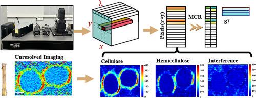
丁香假单胞菌。猕猴桃细菌性溃疡病的病原菌猕猴桃酸菌(actinidiae, Psa)给猕猴桃产业带来了巨大的损失。从细胞和微观层面研究感染过程对制定有效的控制策略具有重要意义。因此,建立合适的化学成像分析方法变得至关重要。共聚焦拉曼微光谱成像(CRMI)与化学计量学相结合,提供了一种直观的方法来可视化和表征健康和感染猕猴桃茎中细胞壁中生物聚合物的时空变化。不同感染茎的拉曼光谱在主成分分析(PCA)中表现出聚类效应,使用支持向量机(SVM)构建的分类模型准确率达到97%。多元曲线分辨率-交替最小二乘法(MCR-ALS)被用于从原始拉曼成像信号中解析光谱矩阵和浓度分布。重建的浓度数据产生高甲基化果胶(HMP)、低甲基化果胶(LMP)、纤维素、半纤维素和木质素的精确分子成像图谱。结果表明,Psa感染3天后,细胞壁中纤维素和HMP含量增加,而半纤维素、木质素和LMP含量变化最小。但在感染5天后,HMP、LMP、纤维素、半纤维素和木质素的含量显著降低,导致细胞壁结构破坏。本研究提出的化学成像方法有望成为研究猕猴桃茎部细菌侵染过程的有效手段。
本文章由计算机程序翻译,如有差异,请以英文原文为准。

Raman imaging-chemometrics analysis of cell wall biopolymer dynamics in Pseudomonas syringae pv. actinidiae-infeicted kiwifruit stems

Raman imaging-chemometrics analysis of cell wall biopolymer dynamics in Pseudomonas syringae pv. actinidiae-infeicted kiwifruit stems
Pseudomonas syringae pv. actinidiae (Psa), the pathogen that causes bacterial canker disease in kiwifruit, has brought about substantial losses to the kiwifruit industry. Investigating the infection process at both the cellular and microscopic levels is of great significance for the formulation of effective control strategies against this disease. Thus, the establishment of appropriate chemical imaging analysis methods becomes essential. Confocal Raman microspectral imaging (CRMI), combined with chemometrics, provides an intuitive means to visualize and characterize the spatiotemporal changes of biopolymers in the cell walls of both healthy and infected kiwifruit stems. Raman spectra of different infected stems exhibit clustering effects in principal component analysis (PCA), and a classification model constructed using support vector machines (SVM) achieves an accuracy of 97 %. Multivariate Curve Resolution-Alternating Least Squares (MCR-ALS) is utilized to resolve spectral matrices and concentration profiles from raw Raman imaging signals. The reconstructed concentration data yields accurate molecular imaging maps of high-methylated pectin (HMP), low-methylated pectin (LMP), cellulose, hemicellulose, and lignin. The results indicate that, three days after Psa infection, the content of cellulose and HMP in the cell wall increases, while the changes in hemicellulose, lignin, and LMP are minimal. However, five days after infection, the contents of HMP, LMP, cellulose, hemicellulose, and lignin decrease significantly, resulting in the disruption of the cell-wall structure. The chemical imaging method proposed in this study shows great promise as an effective means for studying the bacterial infection process in kiwifruit stems at the cellular level.
求助全文
通过发布文献求助,成功后即可免费获取论文全文。 去求助
来源期刊
CiteScore
3.60
自引率
25.00%
发文量
17223
审稿时长
35 days
期刊介绍: Chinese Journal of Analytical Chemistry(CJAC) is an academic journal of analytical chemistry established in 1972 and sponsored by the Chinese Chemical Society and Changchun Institute of Applied Chemistry, Chinese Academy of Sciences. Its objectives are to report the original scientific research achievements and review the recent development of analytical chemistry in all areas. The journal sets up 5 columns including Research Papers, Research Notes, Experimental Technique and Instrument, Review and Progress and Summary Accounts. The journal published monthly in Chinese language. A detailed abstract, keywords and the titles of figures and tables are provided in English, except column of Summary Accounts. Prof. Wang Erkang, an outstanding analytical chemist, academician of Chinese Academy of Sciences & Third World Academy of Sciences, holds the post of the Editor-in-chief.
×
引用
GB/T 7714-2015
复制
MLA
复制
APA
复制
导出至
BibTeX EndNote RefMan NoteFirst NoteExpress
×
提示
您的信息不完整,为了账户安全,请先补充。
现在去补充
×
提示
您因"违规操作"
具体请查看互助需知
我知道了
×
提示
确定
请完成安全验证×
copy
已复制链接
快去分享给好友吧!
我知道了
右上角分享
点击右上角分享
0
联系我们:info@booksci.cn Book学术提供免费学术资源搜索服务,方便国内外学者检索中英文文献。致力于提供最便捷和优质的服务体验。 Copyright © 2023 布克学术 All rights reserved.
京ICP备2023020795号-1
ghs 京公网安备 11010802042870号
Book学术文献互助
Book学术文献互助群
群 号:604180095
Book学术官方微信