Patrick Schindler, Ulrike Grittner, Rebekka Rust, Susanna Asseyer, Judith Bellmann-Strobl, Tanja Schmitz-Hübsch, Michael Scheel, Sven Jarius, Brigitte Wildemann, Markus Reindl, Pascal Benkert, Jens Kuhle, Friedemann Paul, Klemens Ruprecht, Claudia Chien
{"title":"aqp4 - igg阳性NMOSD和MOGAD患者血清GFAP和NfL与脑和上颈MRI体积关系的研究","authors":"Patrick Schindler, Ulrike Grittner, Rebekka Rust, Susanna Asseyer, Judith Bellmann-Strobl, Tanja Schmitz-Hübsch, Michael Scheel, Sven Jarius, Brigitte Wildemann, Markus Reindl, Pascal Benkert, Jens Kuhle, Friedemann Paul, Klemens Ruprecht, Claudia Chien","doi":"10.1177/17562864251345792","DOIUrl":null,"url":null,"abstract":"<p><strong>Background: </strong>Serum glial fibrillary acidic protein (sGFAP) is associated with disease activity in aquaporin-4-immunoglobulin G-seropositive neuromyelitis optica spectrum disorders (AQP4-IgG+NMOSD). Serum neurofilament light chain (sNfL) is a biomarker for neuroaxonal damage. However, the association of sGFAP and sNfL with magnetic resonance imaging (MRI) volumes in AQP4-IgG+NMOSD is unclear.</p><p><strong>Objectives: </strong>To investigate the associations of sGFAP and sNfL with brain MRI volumes in AQP4-IgG+NMOSD.</p><p><strong>Design: </strong>Monocentric, retrospective, observational study.</p><p><strong>Methods: </strong>In 33 clinically stable patients with AQP4-IgG+NMOSD, 17 patients with myelin oligodendrocyte glycoprotein antibody-associated disease (MOGAD), and 15 healthy controls (HC), sGFAP and sNfL were measured at 2 (HC = 1) and 3-Tesla MRIs were obtained at 4 (HC = 1) yearly visits. Associations between biomarkers and MRI metrics were evaluated using linear models.</p><p><strong>Results: </strong>In AQP4-IgG+NMOSD, but not in MOGAD and HC, higher sGFAP was associated with lower hippocampus (β = -2.0 (95% confidence interval: -3.4, -0.7), <i>p</i> = 0.004) and thalamus volumes (β = -2.5 (-4.3, -0.7), <i>p</i> = 0.006) and higher MRI cerebrospinal fluid volume (β = 1.8 (0.7, 3.2), <i>p</i> = 0.01), and, statistically less robust, with lower whole brain (β = -2.3 (-5.3, 0.8), <i>p</i> = 0.15) and gray matter volumes (β = -1.8 (-4.0, 0.4), <i>p</i> = 0.10). Furthermore, higher sGFAP (β = -0.06 (-0.11, -0.002), <i>p</i> = 0.04), but not sNfL (β = -0.02 (-0.08, 0.03), <i>p</i> = 0.38), was associated with percent brain volume change in AQP4-IgG+NMOSD.</p><p><strong>Conclusion: </strong>The specific associations of sGFAP with brain MRI volumes corroborate sGFAP as a biomarker for disease activity in AQP4-IgG+NMOSD.</p>","PeriodicalId":22980,"journal":{"name":"Therapeutic Advances in Neurological Disorders","volume":"18 ","pages":"17562864251345792"},"PeriodicalIF":4.1000,"publicationDate":"2025-07-20","publicationTypes":"Journal Article","fieldsOfStudy":null,"isOpenAccess":false,"openAccessPdf":"https://www.ncbi.nlm.nih.gov/pmc/articles/PMC12277671/pdf/","citationCount":"0","resultStr":"{\"title\":\"Investigation of the association of serum GFAP and NfL with brain and upper cervical MRI volumes in AQP4-IgG-positive NMOSD and MOGAD.\",\"authors\":\"Patrick Schindler, Ulrike Grittner, Rebekka Rust, Susanna Asseyer, Judith Bellmann-Strobl, Tanja Schmitz-Hübsch, Michael Scheel, Sven Jarius, Brigitte Wildemann, Markus Reindl, Pascal Benkert, Jens Kuhle, Friedemann Paul, Klemens Ruprecht, Claudia Chien\",\"doi\":\"10.1177/17562864251345792\",\"DOIUrl\":null,\"url\":null,\"abstract\":\"<p><strong>Background: </strong>Serum glial fibrillary acidic protein (sGFAP) is associated with disease activity in aquaporin-4-immunoglobulin G-seropositive neuromyelitis optica spectrum disorders (AQP4-IgG+NMOSD). Serum neurofilament light chain (sNfL) is a biomarker for neuroaxonal damage. However, the association of sGFAP and sNfL with magnetic resonance imaging (MRI) volumes in AQP4-IgG+NMOSD is unclear.</p><p><strong>Objectives: </strong>To investigate the associations of sGFAP and sNfL with brain MRI volumes in AQP4-IgG+NMOSD.</p><p><strong>Design: </strong>Monocentric, retrospective, observational study.</p><p><strong>Methods: </strong>In 33 clinically stable patients with AQP4-IgG+NMOSD, 17 patients with myelin oligodendrocyte glycoprotein antibody-associated disease (MOGAD), and 15 healthy controls (HC), sGFAP and sNfL were measured at 2 (HC = 1) and 3-Tesla MRIs were obtained at 4 (HC = 1) yearly visits. Associations between biomarkers and MRI metrics were evaluated using linear models.</p><p><strong>Results: </strong>In AQP4-IgG+NMOSD, but not in MOGAD and HC, higher sGFAP was associated with lower hippocampus (β = -2.0 (95% confidence interval: -3.4, -0.7), <i>p</i> = 0.004) and thalamus volumes (β = -2.5 (-4.3, -0.7), <i>p</i> = 0.006) and higher MRI cerebrospinal fluid volume (β = 1.8 (0.7, 3.2), <i>p</i> = 0.01), and, statistically less robust, with lower whole brain (β = -2.3 (-5.3, 0.8), <i>p</i> = 0.15) and gray matter volumes (β = -1.8 (-4.0, 0.4), <i>p</i> = 0.10). Furthermore, higher sGFAP (β = -0.06 (-0.11, -0.002), <i>p</i> = 0.04), but not sNfL (β = -0.02 (-0.08, 0.03), <i>p</i> = 0.38), was associated with percent brain volume change in AQP4-IgG+NMOSD.</p><p><strong>Conclusion: </strong>The specific associations of sGFAP with brain MRI volumes corroborate sGFAP as a biomarker for disease activity in AQP4-IgG+NMOSD.</p>\",\"PeriodicalId\":22980,\"journal\":{\"name\":\"Therapeutic Advances in Neurological Disorders\",\"volume\":\"18 \",\"pages\":\"17562864251345792\"},\"PeriodicalIF\":4.1000,\"publicationDate\":\"2025-07-20\",\"publicationTypes\":\"Journal Article\",\"fieldsOfStudy\":null,\"isOpenAccess\":false,\"openAccessPdf\":\"https://www.ncbi.nlm.nih.gov/pmc/articles/PMC12277671/pdf/\",\"citationCount\":\"0\",\"resultStr\":null,\"platform\":\"Semanticscholar\",\"paperid\":null,\"PeriodicalName\":\"Therapeutic Advances in Neurological Disorders\",\"FirstCategoryId\":\"3\",\"ListUrlMain\":\"https://doi.org/10.1177/17562864251345792\",\"RegionNum\":2,\"RegionCategory\":\"医学\",\"ArticlePicture\":[],\"TitleCN\":null,\"AbstractTextCN\":null,\"PMCID\":null,\"EPubDate\":\"2025/1/1 0:00:00\",\"PubModel\":\"eCollection\",\"JCR\":\"Q1\",\"JCRName\":\"CLINICAL NEUROLOGY\",\"Score\":null,\"Total\":0}","platform":"Semanticscholar","paperid":null,"PeriodicalName":"Therapeutic Advances in Neurological Disorders","FirstCategoryId":"3","ListUrlMain":"https://doi.org/10.1177/17562864251345792","RegionNum":2,"RegionCategory":"医学","ArticlePicture":[],"TitleCN":null,"AbstractTextCN":null,"PMCID":null,"EPubDate":"2025/1/1 0:00:00","PubModel":"eCollection","JCR":"Q1","JCRName":"CLINICAL NEUROLOGY","Score":null,"Total":0}
Investigation of the association of serum GFAP and NfL with brain and upper cervical MRI volumes in AQP4-IgG-positive NMOSD and MOGAD.
Background: Serum glial fibrillary acidic protein (sGFAP) is associated with disease activity in aquaporin-4-immunoglobulin G-seropositive neuromyelitis optica spectrum disorders (AQP4-IgG+NMOSD). Serum neurofilament light chain (sNfL) is a biomarker for neuroaxonal damage. However, the association of sGFAP and sNfL with magnetic resonance imaging (MRI) volumes in AQP4-IgG+NMOSD is unclear.
Objectives: To investigate the associations of sGFAP and sNfL with brain MRI volumes in AQP4-IgG+NMOSD.
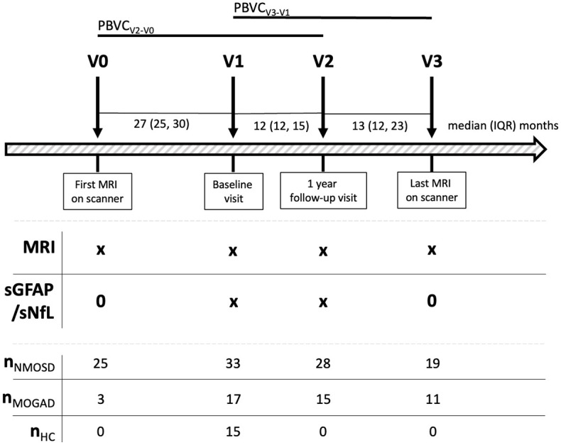
Methods: In 33 clinically stable patients with AQP4-IgG+NMOSD, 17 patients with myelin oligodendrocyte glycoprotein antibody-associated disease (MOGAD), and 15 healthy controls (HC), sGFAP and sNfL were measured at 2 (HC = 1) and 3-Tesla MRIs were obtained at 4 (HC = 1) yearly visits. Associations between biomarkers and MRI metrics were evaluated using linear models.
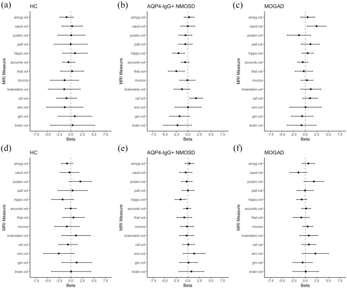
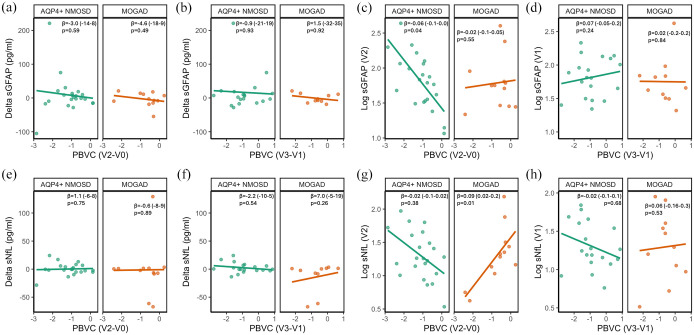
Results: In AQP4-IgG+NMOSD, but not in MOGAD and HC, higher sGFAP was associated with lower hippocampus (β = -2.0 (95% confidence interval: -3.4, -0.7), p = 0.004) and thalamus volumes (β = -2.5 (-4.3, -0.7), p = 0.006) and higher MRI cerebrospinal fluid volume (β = 1.8 (0.7, 3.2), p = 0.01), and, statistically less robust, with lower whole brain (β = -2.3 (-5.3, 0.8), p = 0.15) and gray matter volumes (β = -1.8 (-4.0, 0.4), p = 0.10). Furthermore, higher sGFAP (β = -0.06 (-0.11, -0.002), p = 0.04), but not sNfL (β = -0.02 (-0.08, 0.03), p = 0.38), was associated with percent brain volume change in AQP4-IgG+NMOSD.
Conclusion: The specific associations of sGFAP with brain MRI volumes corroborate sGFAP as a biomarker for disease activity in AQP4-IgG+NMOSD.
期刊介绍:
Therapeutic Advances in Neurological Disorders is a peer-reviewed, open access journal delivering the highest quality articles, reviews, and scholarly comment on pioneering efforts and innovative studies across all areas of neurology. The journal has a strong clinical and pharmacological focus and is aimed at clinicians and researchers in neurology, providing a forum in print and online for publishing the highest quality articles in this area.
 求助内容:
求助内容: 应助结果提醒方式:


