Nicole Dausend, Evan J Cosgrove, Paula R Giaretta, Philip H Kass, Valerie Freiche, Sina Marsilio
{"title":"慢性肠病猫粘膜纤维化的定量评估及其与疾病严重程度和预后的相关性","authors":"Nicole Dausend, Evan J Cosgrove, Paula R Giaretta, Philip H Kass, Valerie Freiche, Sina Marsilio","doi":"10.1177/1098612X251338174","DOIUrl":null,"url":null,"abstract":"<p><p>ObjectivesThis study aimed to develop a quantitative scoring method for the evaluation of mucosal fibrosis (MF) and assess its correlation with World Small Animal Veterinary Association scores, clinical abnormalities, disease severity, diagnosis and outcome in cats with lymphoplasmacytic enteritis (LPE) or low-grade intestinal T-cell lymphoma (LGITL).MethodsFormalin-fixed, paraffin-embedded small intestinal biopsy specimens from 13 cats with LPE and 14 cats with LGITL were included. MF was quantitatively measured in three separate areas (villi, apical crypts [ACs] and basal crypts) using an image processing program. The fractional fibrotic area (%FFA) was calculated based on the mean fibrosis scores in five representative fields. MF was also scored by a single board-certified pathologist on sequential slides stained with either hematoxylin and eosin (H&E) or Masson's trichrome (MT) on a four-point scale in the areas described above. Statistical analysis was performed to assess the correlation between clinical and pathological variables, diagnosis and outcome.ResultsThe %FFA scores correlated well with fibrosis scores on MT stains (<i>r</i> = 0.52, <i>P</i> = 0.01) but did not correlate with H&E stains (<i>r</i> = 0.29, <i>P</i> = 0.14). The %FFA in the villi and AC area was negatively correlated with a modified Feline Chronic Enteropathy Activity Index in cats with LGITL (<i>r</i> = -0.57, <i>P</i> = 0.04). A histopathologic diagnosis of LPE showed a weak correlation with MF in the AC area (<i>r</i> = 0.38, <i>P</i> = 0.05). The survival time of cats with chronic enteropathy (CE) was weakly negatively correlated with MF (<i>r</i> = -0.38, <i>P</i> = 0.05).Conclusions and relevanceMF is more effectively assessed using MT staining compared with H&E staining alone. Increased MF in the AC region may indicate a diagnosis of LPE. Although increased MF did not correlate with increased disease activity, it appears to be a negative prognostic factor for survival in cats with CE.</p>","PeriodicalId":15851,"journal":{"name":"Journal of Feline Medicine and Surgery","volume":"27 7","pages":"1098612X251338174"},"PeriodicalIF":2.1000,"publicationDate":"2025-07-01","publicationTypes":"Journal Article","fieldsOfStudy":null,"isOpenAccess":false,"openAccessPdf":"https://www.ncbi.nlm.nih.gov/pmc/articles/PMC12290242/pdf/","citationCount":"0","resultStr":"{\"title\":\"Quantitative assessment of mucosal fibrosis and its correlation with disease severity and outcome in cats with chronic enteropathy.\",\"authors\":\"Nicole Dausend, Evan J Cosgrove, Paula R Giaretta, Philip H Kass, Valerie Freiche, Sina Marsilio\",\"doi\":\"10.1177/1098612X251338174\",\"DOIUrl\":null,\"url\":null,\"abstract\":\"<p><p>ObjectivesThis study aimed to develop a quantitative scoring method for the evaluation of mucosal fibrosis (MF) and assess its correlation with World Small Animal Veterinary Association scores, clinical abnormalities, disease severity, diagnosis and outcome in cats with lymphoplasmacytic enteritis (LPE) or low-grade intestinal T-cell lymphoma (LGITL).MethodsFormalin-fixed, paraffin-embedded small intestinal biopsy specimens from 13 cats with LPE and 14 cats with LGITL were included. MF was quantitatively measured in three separate areas (villi, apical crypts [ACs] and basal crypts) using an image processing program. The fractional fibrotic area (%FFA) was calculated based on the mean fibrosis scores in five representative fields. MF was also scored by a single board-certified pathologist on sequential slides stained with either hematoxylin and eosin (H&E) or Masson's trichrome (MT) on a four-point scale in the areas described above. Statistical analysis was performed to assess the correlation between clinical and pathological variables, diagnosis and outcome.ResultsThe %FFA scores correlated well with fibrosis scores on MT stains (<i>r</i> = 0.52, <i>P</i> = 0.01) but did not correlate with H&E stains (<i>r</i> = 0.29, <i>P</i> = 0.14). The %FFA in the villi and AC area was negatively correlated with a modified Feline Chronic Enteropathy Activity Index in cats with LGITL (<i>r</i> = -0.57, <i>P</i> = 0.04). A histopathologic diagnosis of LPE showed a weak correlation with MF in the AC area (<i>r</i> = 0.38, <i>P</i> = 0.05). The survival time of cats with chronic enteropathy (CE) was weakly negatively correlated with MF (<i>r</i> = -0.38, <i>P</i> = 0.05).Conclusions and relevanceMF is more effectively assessed using MT staining compared with H&E staining alone. Increased MF in the AC region may indicate a diagnosis of LPE. Although increased MF did not correlate with increased disease activity, it appears to be a negative prognostic factor for survival in cats with CE.</p>\",\"PeriodicalId\":15851,\"journal\":{\"name\":\"Journal of Feline Medicine and Surgery\",\"volume\":\"27 7\",\"pages\":\"1098612X251338174\"},\"PeriodicalIF\":2.1000,\"publicationDate\":\"2025-07-01\",\"publicationTypes\":\"Journal Article\",\"fieldsOfStudy\":null,\"isOpenAccess\":false,\"openAccessPdf\":\"https://www.ncbi.nlm.nih.gov/pmc/articles/PMC12290242/pdf/\",\"citationCount\":\"0\",\"resultStr\":null,\"platform\":\"Semanticscholar\",\"paperid\":null,\"PeriodicalName\":\"Journal of Feline Medicine and Surgery\",\"FirstCategoryId\":\"97\",\"ListUrlMain\":\"https://doi.org/10.1177/1098612X251338174\",\"RegionNum\":2,\"RegionCategory\":\"农林科学\",\"ArticlePicture\":[],\"TitleCN\":null,\"AbstractTextCN\":null,\"PMCID\":null,\"EPubDate\":\"2025/7/22 0:00:00\",\"PubModel\":\"Epub\",\"JCR\":\"Q2\",\"JCRName\":\"VETERINARY SCIENCES\",\"Score\":null,\"Total\":0}","platform":"Semanticscholar","paperid":null,"PeriodicalName":"Journal of Feline Medicine and Surgery","FirstCategoryId":"97","ListUrlMain":"https://doi.org/10.1177/1098612X251338174","RegionNum":2,"RegionCategory":"农林科学","ArticlePicture":[],"TitleCN":null,"AbstractTextCN":null,"PMCID":null,"EPubDate":"2025/7/22 0:00:00","PubModel":"Epub","JCR":"Q2","JCRName":"VETERINARY SCIENCES","Score":null,"Total":0}
引用次数: 0
摘要
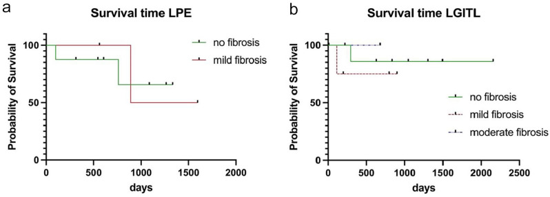
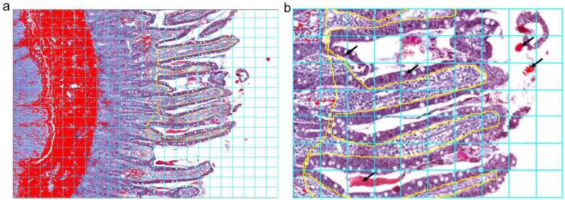
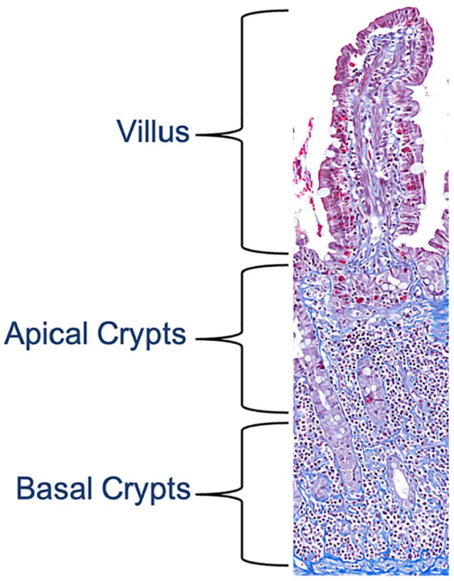
本研究旨在建立一种定量评分方法来评估猫淋巴浆细胞性肠炎(LPE)或低级别肠t细胞淋巴瘤(llgl)的黏膜纤维化(MF),并评估其与世界小动物兽医协会评分、临床异常、疾病严重程度、诊断和预后的相关性。方法对13只LPE猫和14只llgl猫进行福尔马林固定、石蜡包埋的小肠活检标本。使用图像处理程序定量测量三个不同区域(绒毛、根尖隐窝和基底隐窝)的MF。分数纤维化面积(%FFA)根据五个代表性领域的平均纤维化评分计算。MF也由一个委员会认证的病理学家在连续的载片上用苏木精和伊红(H&E)或马森三色(MT)在上述区域以四分制进行评分。通过统计学分析评估临床和病理变量、诊断和预后之间的相关性。结果%FFA评分与MT染色纤维化评分相关性较好(r = 0.52, P = 0.01),与H&E染色评分相关性不高(r = 0.29, P = 0.14)。lgil猫的绒毛和AC区FFA百分比与修正后的猫慢性肠病活动指数呈负相关(r = -0.57, P = 0.04)。组织病理学诊断LPE与AC区MF呈弱相关性(r = 0.38, P = 0.05)。慢性肠病(CE)猫的生存时间与MF呈弱负相关(r = -0.38, P = 0.05)。结论及相关性与单独H&E染色相比,MT染色能更有效地评估emf。AC区MF增高可能提示LPE的诊断。虽然MF增加与疾病活动性增加无关,但它似乎是CE猫生存的负面预后因素。
Quantitative assessment of mucosal fibrosis and its correlation with disease severity and outcome in cats with chronic enteropathy.
ObjectivesThis study aimed to develop a quantitative scoring method for the evaluation of mucosal fibrosis (MF) and assess its correlation with World Small Animal Veterinary Association scores, clinical abnormalities, disease severity, diagnosis and outcome in cats with lymphoplasmacytic enteritis (LPE) or low-grade intestinal T-cell lymphoma (LGITL).MethodsFormalin-fixed, paraffin-embedded small intestinal biopsy specimens from 13 cats with LPE and 14 cats with LGITL were included. MF was quantitatively measured in three separate areas (villi, apical crypts [ACs] and basal crypts) using an image processing program. The fractional fibrotic area (%FFA) was calculated based on the mean fibrosis scores in five representative fields. MF was also scored by a single board-certified pathologist on sequential slides stained with either hematoxylin and eosin (H&E) or Masson's trichrome (MT) on a four-point scale in the areas described above. Statistical analysis was performed to assess the correlation between clinical and pathological variables, diagnosis and outcome.ResultsThe %FFA scores correlated well with fibrosis scores on MT stains (r = 0.52, P = 0.01) but did not correlate with H&E stains (r = 0.29, P = 0.14). The %FFA in the villi and AC area was negatively correlated with a modified Feline Chronic Enteropathy Activity Index in cats with LGITL (r = -0.57, P = 0.04). A histopathologic diagnosis of LPE showed a weak correlation with MF in the AC area (r = 0.38, P = 0.05). The survival time of cats with chronic enteropathy (CE) was weakly negatively correlated with MF (r = -0.38, P = 0.05).Conclusions and relevanceMF is more effectively assessed using MT staining compared with H&E staining alone. Increased MF in the AC region may indicate a diagnosis of LPE. Although increased MF did not correlate with increased disease activity, it appears to be a negative prognostic factor for survival in cats with CE.
期刊介绍:
JFMS is an international, peer-reviewed journal aimed at both practitioners and researchers with an interest in the clinical veterinary healthcare of domestic cats. The journal is published monthly in two formats: ‘Classic’ editions containing high-quality original papers on all aspects of feline medicine and surgery, including basic research relevant to clinical practice; and dedicated ‘Clinical Practice’ editions primarily containing opinionated review articles providing state-of-the-art information for feline clinicians, along with other relevant articles such as consensus guidelines.
 求助内容:
求助内容: 应助结果提醒方式:


