Jeffrey G Malins, D M Anisuzzaman, John I Jackson, Eunjung Lee, Jwan A Naser, Jared G Bird, Paul A Friedman, Christie C Ngo, Jae K Oh, Gal Tsaban, Patricia A Pellikka, Jeremy J Thaden, Francisco Lopez-Jimenez, Zachi I Attia, Sorin V Pislaru, Garvan C Kane
{"title":"用于经胸超声心动图和手持式心脏超声左心室增大分类的深度学习模型。","authors":"Jeffrey G Malins, D M Anisuzzaman, John I Jackson, Eunjung Lee, Jwan A Naser, Jared G Bird, Paul A Friedman, Christie C Ngo, Jae K Oh, Gal Tsaban, Patricia A Pellikka, Jeremy J Thaden, Francisco Lopez-Jimenez, Zachi I Attia, Sorin V Pislaru, Garvan C Kane","doi":"10.1093/ehjimp/qyaf049","DOIUrl":null,"url":null,"abstract":"<p><strong>Aims: </strong>To develop a deep learning model that: (i) utilizes transthoracic echocardiography (TTE) clips to detect left ventricular (LV) enlargement without being provided information regarding a patient's sex and body size; and (ii) can be accurately applied to clips acquired using either standard comprehensive TTE or handheld cardiac ultrasound (HCU).</p><p><strong>Methods and results: </strong>Using retrospective TTE data (training: 8722 patients; internal validation: 468 patients), we developed a deep learning model that estimates a patient's end-diastolic LV volume (indexed to body surface area and normalized across the sexes), and then thresholds this estimate to perform the following classifications: (1) normally sized LV vs. ≥ mild LV enlargement; (2) normal/mildly enlarged LV vs. ≥ moderate LV enlargement. For retrospective datasets, the model showed strong performance in TTE across three geographically distinct locations (Minnesota and Wisconsin: 1082 patients, AUC = 0.925 and 0.953 for classifications 1 and 2, respectively; Arizona: 1475 patients, AUC = 0.935 and 0.969; and Florida: 1481 patients, AUC = 0.934 and 0.970). Additionally, performance was strong for both TTE and HCU clips collected from a prospective cohort of 410 patients who underwent HCU immediately following TTE (TTE: AUC = 0.925 and 0.971; HCU: AUC = 0.874 and 0.902, for classifications 1 and 2, respectively).</p><p><strong>Conclusion: </strong>An automated deep learning model applied to TTE or HCU images accurately categorizes LV volumes. These results lay a foundation for future work aimed at optimizing clinical outcomes for heart failure patients by enabling early detection of LV enlargement across various point-of-care settings.</p>","PeriodicalId":94317,"journal":{"name":"European heart journal. Imaging methods and practice","volume":"3 3","pages":"qyaf049"},"PeriodicalIF":0.0000,"publicationDate":"2025-05-09","publicationTypes":"Journal Article","fieldsOfStudy":null,"isOpenAccess":false,"openAccessPdf":"https://www.ncbi.nlm.nih.gov/pmc/articles/PMC12275095/pdf/","citationCount":"0","resultStr":"{\"title\":\"A deep learning model for classifying left ventricular enlargement for both transthoracic echocardiograms and handheld cardiac ultrasound.\",\"authors\":\"Jeffrey G Malins, D M Anisuzzaman, John I Jackson, Eunjung Lee, Jwan A Naser, Jared G Bird, Paul A Friedman, Christie C Ngo, Jae K Oh, Gal Tsaban, Patricia A Pellikka, Jeremy J Thaden, Francisco Lopez-Jimenez, Zachi I Attia, Sorin V Pislaru, Garvan C Kane\",\"doi\":\"10.1093/ehjimp/qyaf049\",\"DOIUrl\":null,\"url\":null,\"abstract\":\"<p><strong>Aims: </strong>To develop a deep learning model that: (i) utilizes transthoracic echocardiography (TTE) clips to detect left ventricular (LV) enlargement without being provided information regarding a patient's sex and body size; and (ii) can be accurately applied to clips acquired using either standard comprehensive TTE or handheld cardiac ultrasound (HCU).</p><p><strong>Methods and results: </strong>Using retrospective TTE data (training: 8722 patients; internal validation: 468 patients), we developed a deep learning model that estimates a patient's end-diastolic LV volume (indexed to body surface area and normalized across the sexes), and then thresholds this estimate to perform the following classifications: (1) normally sized LV vs. ≥ mild LV enlargement; (2) normal/mildly enlarged LV vs. ≥ moderate LV enlargement. For retrospective datasets, the model showed strong performance in TTE across three geographically distinct locations (Minnesota and Wisconsin: 1082 patients, AUC = 0.925 and 0.953 for classifications 1 and 2, respectively; Arizona: 1475 patients, AUC = 0.935 and 0.969; and Florida: 1481 patients, AUC = 0.934 and 0.970). Additionally, performance was strong for both TTE and HCU clips collected from a prospective cohort of 410 patients who underwent HCU immediately following TTE (TTE: AUC = 0.925 and 0.971; HCU: AUC = 0.874 and 0.902, for classifications 1 and 2, respectively).</p><p><strong>Conclusion: </strong>An automated deep learning model applied to TTE or HCU images accurately categorizes LV volumes. These results lay a foundation for future work aimed at optimizing clinical outcomes for heart failure patients by enabling early detection of LV enlargement across various point-of-care settings.</p>\",\"PeriodicalId\":94317,\"journal\":{\"name\":\"European heart journal. Imaging methods and practice\",\"volume\":\"3 3\",\"pages\":\"qyaf049\"},\"PeriodicalIF\":0.0000,\"publicationDate\":\"2025-05-09\",\"publicationTypes\":\"Journal Article\",\"fieldsOfStudy\":null,\"isOpenAccess\":false,\"openAccessPdf\":\"https://www.ncbi.nlm.nih.gov/pmc/articles/PMC12275095/pdf/\",\"citationCount\":\"0\",\"resultStr\":null,\"platform\":\"Semanticscholar\",\"paperid\":null,\"PeriodicalName\":\"European heart journal. Imaging methods and practice\",\"FirstCategoryId\":\"1085\",\"ListUrlMain\":\"https://doi.org/10.1093/ehjimp/qyaf049\",\"RegionNum\":0,\"RegionCategory\":null,\"ArticlePicture\":[],\"TitleCN\":null,\"AbstractTextCN\":null,\"PMCID\":null,\"EPubDate\":\"2024/8/1 0:00:00\",\"PubModel\":\"eCollection\",\"JCR\":\"\",\"JCRName\":\"\",\"Score\":null,\"Total\":0}","platform":"Semanticscholar","paperid":null,"PeriodicalName":"European heart journal. Imaging methods and practice","FirstCategoryId":"1085","ListUrlMain":"https://doi.org/10.1093/ehjimp/qyaf049","RegionNum":0,"RegionCategory":null,"ArticlePicture":[],"TitleCN":null,"AbstractTextCN":null,"PMCID":null,"EPubDate":"2024/8/1 0:00:00","PubModel":"eCollection","JCR":"","JCRName":"","Score":null,"Total":0}
A deep learning model for classifying left ventricular enlargement for both transthoracic echocardiograms and handheld cardiac ultrasound.
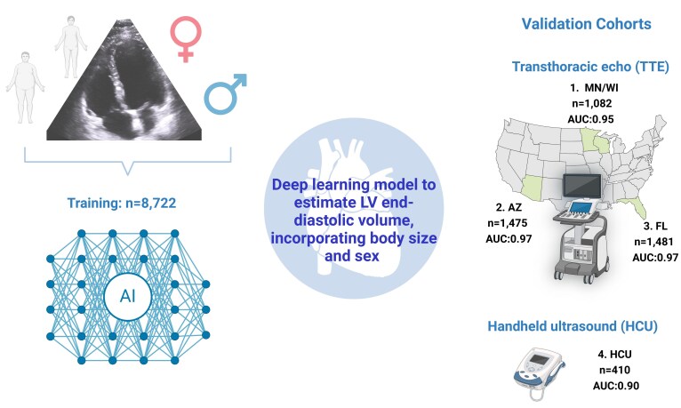
Aims: To develop a deep learning model that: (i) utilizes transthoracic echocardiography (TTE) clips to detect left ventricular (LV) enlargement without being provided information regarding a patient's sex and body size; and (ii) can be accurately applied to clips acquired using either standard comprehensive TTE or handheld cardiac ultrasound (HCU).
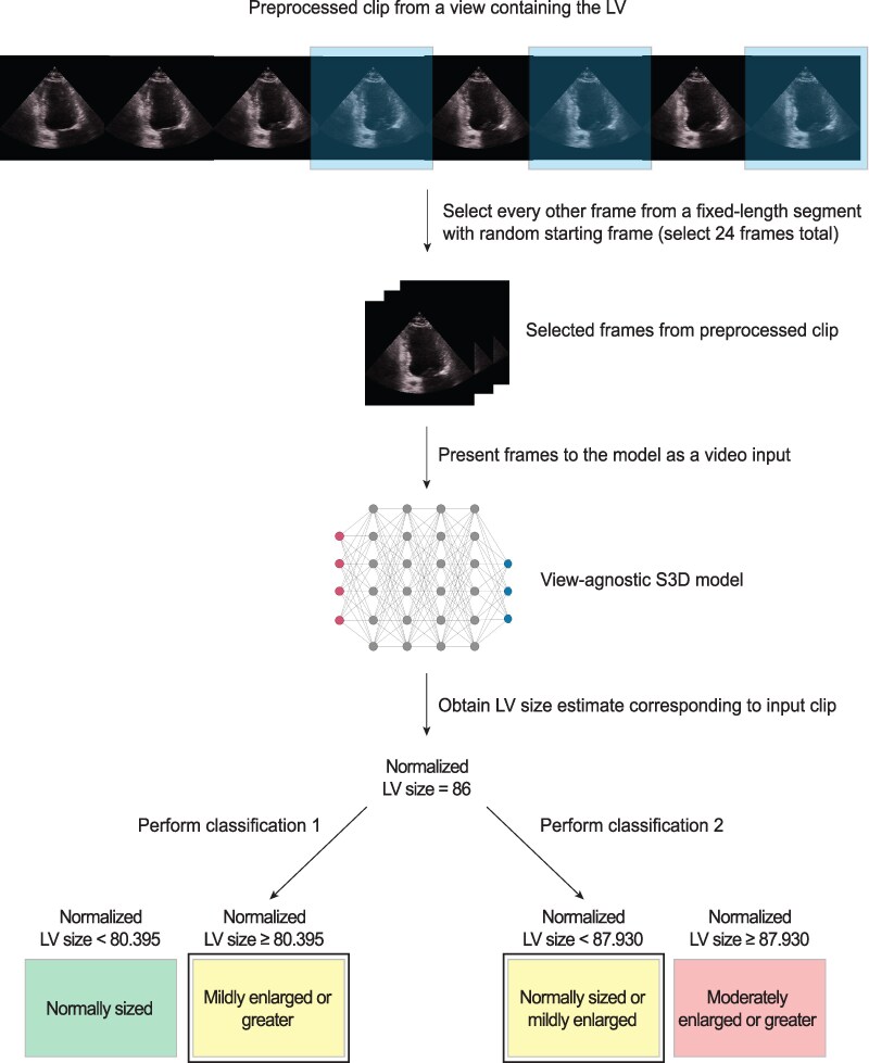
Methods and results: Using retrospective TTE data (training: 8722 patients; internal validation: 468 patients), we developed a deep learning model that estimates a patient's end-diastolic LV volume (indexed to body surface area and normalized across the sexes), and then thresholds this estimate to perform the following classifications: (1) normally sized LV vs. ≥ mild LV enlargement; (2) normal/mildly enlarged LV vs. ≥ moderate LV enlargement. For retrospective datasets, the model showed strong performance in TTE across three geographically distinct locations (Minnesota and Wisconsin: 1082 patients, AUC = 0.925 and 0.953 for classifications 1 and 2, respectively; Arizona: 1475 patients, AUC = 0.935 and 0.969; and Florida: 1481 patients, AUC = 0.934 and 0.970). Additionally, performance was strong for both TTE and HCU clips collected from a prospective cohort of 410 patients who underwent HCU immediately following TTE (TTE: AUC = 0.925 and 0.971; HCU: AUC = 0.874 and 0.902, for classifications 1 and 2, respectively).
Conclusion: An automated deep learning model applied to TTE or HCU images accurately categorizes LV volumes. These results lay a foundation for future work aimed at optimizing clinical outcomes for heart failure patients by enabling early detection of LV enlargement across various point-of-care settings.


 求助内容:
求助内容: 应助结果提醒方式:


