Maciej Janeczek, Karolina Goździewska-Harłajczuk, Agata Małyszek, Ludwika Hrabska, Joanna Klećkowska-Nawrot
{"title":"科莫多龙(Varanus komodoensis)腭襞唾液腺和下颌毒腺的组织学和组织化学特征。","authors":"Maciej Janeczek, Karolina Goździewska-Harłajczuk, Agata Małyszek, Ludwika Hrabska, Joanna Klećkowska-Nawrot","doi":"10.1007/s11259-025-10825-6","DOIUrl":null,"url":null,"abstract":"<p><p>The Komodo dragon (Varanus komodoensis) is the largest living lizard whose hunting strategy allows it to attack large animals. It inflicts serious damage to its prey with specially developed teeth, called ziphodonts. Although the mandibular venom gland system was previously detected in the Komodo dragon, there is still a lack of its histochemical analysis. Thus, the objective of the current study was a detailed description of the mandibular venom gland of this species. In addition, a histological examination of the salivary glands of the palatine fold was performed. The research material was collected post-mortem from the captive adult female Komodo dragon. Hematoxylin and eosin and Masson-Goldner trichrome staining methods were used for the histological analysis of the glands, while periodic acid-Schiff, alcian blue pH 1.0, alcian blue pH 2.5, alcian blue pH 2.5 PAS and Hale's dialysed iron methods were included for the histochemical study. The venom gland was composed of distinctly marked and very numerous individual lobes surrounded by dense, irregularly structured, highly developed connective tissue that forms the interlobar septa. The salivary glands of the palatine fold were surrounded by a thick connective tissue capsule made of dense, irregularly structured connective tissue. The single ducts of the mandibular venom gland open into the sheaths surrounding the consecutive teeth. The presence of numerous muscle cells in the stroma of the venom gland between its lobes may indicate their participation in the emptying of the vesicles of their secretion.</p>","PeriodicalId":23690,"journal":{"name":"Veterinary Research Communications","volume":"49 5","pages":"260"},"PeriodicalIF":2.0000,"publicationDate":"2025-07-21","publicationTypes":"Journal Article","fieldsOfStudy":null,"isOpenAccess":false,"openAccessPdf":"https://www.ncbi.nlm.nih.gov/pmc/articles/PMC12279577/pdf/","citationCount":"0","resultStr":"{\"title\":\"Histological and histochemical characterisation of the salivary glands of the palatine fold and the mandibular venom gland of the Komodo dragon (Varanus komodoensis).\",\"authors\":\"Maciej Janeczek, Karolina Goździewska-Harłajczuk, Agata Małyszek, Ludwika Hrabska, Joanna Klećkowska-Nawrot\",\"doi\":\"10.1007/s11259-025-10825-6\",\"DOIUrl\":null,\"url\":null,\"abstract\":\"<p><p>The Komodo dragon (Varanus komodoensis) is the largest living lizard whose hunting strategy allows it to attack large animals. It inflicts serious damage to its prey with specially developed teeth, called ziphodonts. Although the mandibular venom gland system was previously detected in the Komodo dragon, there is still a lack of its histochemical analysis. Thus, the objective of the current study was a detailed description of the mandibular venom gland of this species. In addition, a histological examination of the salivary glands of the palatine fold was performed. The research material was collected post-mortem from the captive adult female Komodo dragon. Hematoxylin and eosin and Masson-Goldner trichrome staining methods were used for the histological analysis of the glands, while periodic acid-Schiff, alcian blue pH 1.0, alcian blue pH 2.5, alcian blue pH 2.5 PAS and Hale's dialysed iron methods were included for the histochemical study. The venom gland was composed of distinctly marked and very numerous individual lobes surrounded by dense, irregularly structured, highly developed connective tissue that forms the interlobar septa. The salivary glands of the palatine fold were surrounded by a thick connective tissue capsule made of dense, irregularly structured connective tissue. The single ducts of the mandibular venom gland open into the sheaths surrounding the consecutive teeth. The presence of numerous muscle cells in the stroma of the venom gland between its lobes may indicate their participation in the emptying of the vesicles of their secretion.</p>\",\"PeriodicalId\":23690,\"journal\":{\"name\":\"Veterinary Research Communications\",\"volume\":\"49 5\",\"pages\":\"260\"},\"PeriodicalIF\":2.0000,\"publicationDate\":\"2025-07-21\",\"publicationTypes\":\"Journal Article\",\"fieldsOfStudy\":null,\"isOpenAccess\":false,\"openAccessPdf\":\"https://www.ncbi.nlm.nih.gov/pmc/articles/PMC12279577/pdf/\",\"citationCount\":\"0\",\"resultStr\":null,\"platform\":\"Semanticscholar\",\"paperid\":null,\"PeriodicalName\":\"Veterinary Research Communications\",\"FirstCategoryId\":\"97\",\"ListUrlMain\":\"https://doi.org/10.1007/s11259-025-10825-6\",\"RegionNum\":3,\"RegionCategory\":\"农林科学\",\"ArticlePicture\":[],\"TitleCN\":null,\"AbstractTextCN\":null,\"PMCID\":null,\"EPubDate\":\"\",\"PubModel\":\"\",\"JCR\":\"Q2\",\"JCRName\":\"VETERINARY SCIENCES\",\"Score\":null,\"Total\":0}","platform":"Semanticscholar","paperid":null,"PeriodicalName":"Veterinary Research Communications","FirstCategoryId":"97","ListUrlMain":"https://doi.org/10.1007/s11259-025-10825-6","RegionNum":3,"RegionCategory":"农林科学","ArticlePicture":[],"TitleCN":null,"AbstractTextCN":null,"PMCID":null,"EPubDate":"","PubModel":"","JCR":"Q2","JCRName":"VETERINARY SCIENCES","Score":null,"Total":0}
Histological and histochemical characterisation of the salivary glands of the palatine fold and the mandibular venom gland of the Komodo dragon (Varanus komodoensis).
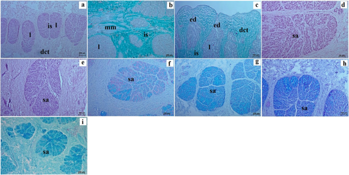
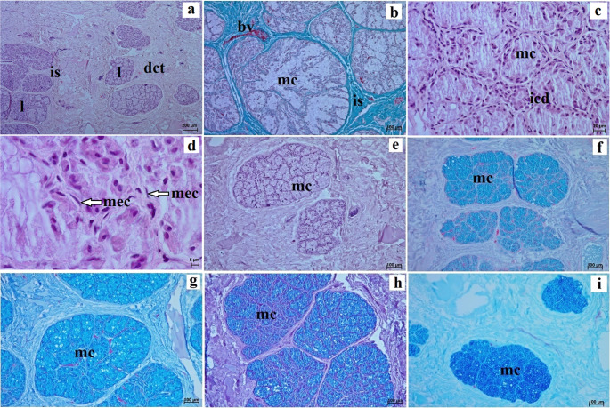
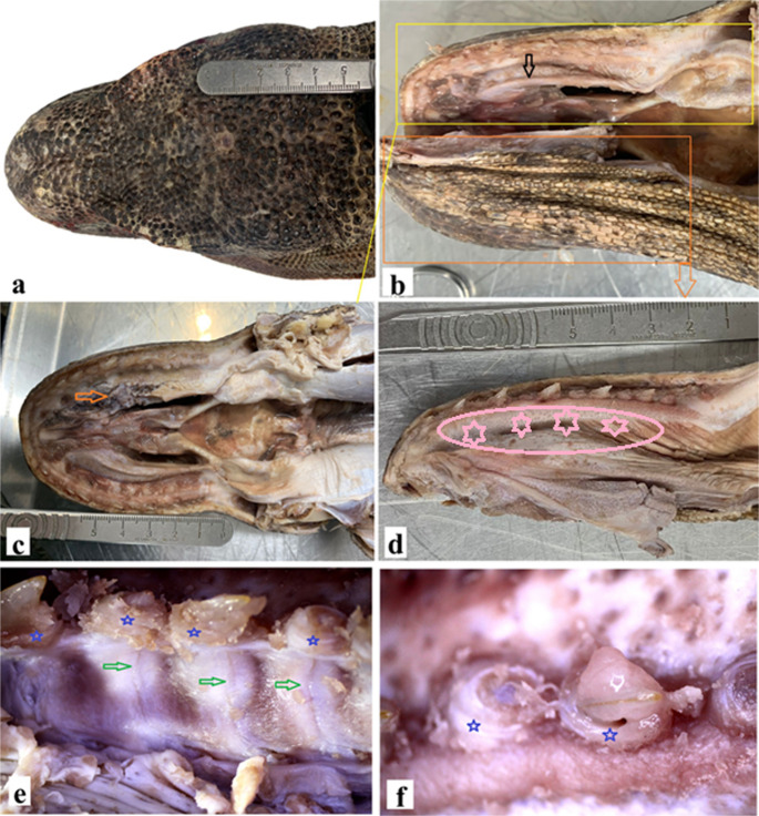
The Komodo dragon (Varanus komodoensis) is the largest living lizard whose hunting strategy allows it to attack large animals. It inflicts serious damage to its prey with specially developed teeth, called ziphodonts. Although the mandibular venom gland system was previously detected in the Komodo dragon, there is still a lack of its histochemical analysis. Thus, the objective of the current study was a detailed description of the mandibular venom gland of this species. In addition, a histological examination of the salivary glands of the palatine fold was performed. The research material was collected post-mortem from the captive adult female Komodo dragon. Hematoxylin and eosin and Masson-Goldner trichrome staining methods were used for the histological analysis of the glands, while periodic acid-Schiff, alcian blue pH 1.0, alcian blue pH 2.5, alcian blue pH 2.5 PAS and Hale's dialysed iron methods were included for the histochemical study. The venom gland was composed of distinctly marked and very numerous individual lobes surrounded by dense, irregularly structured, highly developed connective tissue that forms the interlobar septa. The salivary glands of the palatine fold were surrounded by a thick connective tissue capsule made of dense, irregularly structured connective tissue. The single ducts of the mandibular venom gland open into the sheaths surrounding the consecutive teeth. The presence of numerous muscle cells in the stroma of the venom gland between its lobes may indicate their participation in the emptying of the vesicles of their secretion.
期刊介绍:
Veterinary Research Communications publishes fully refereed research articles and topical reviews on all aspects of the veterinary sciences. Interdisciplinary articles are particularly encouraged, as are well argued reviews, even if they are somewhat controversial.
The journal is an appropriate medium in which to publish new methods, newly described diseases and new pathological findings, as these are applied to animals. The material should be of international rather than local interest. As it deliberately seeks a wide coverage, Veterinary Research Communications provides its readers with a means of keeping abreast of current developments in the entire field of veterinary science.
 求助内容:
求助内容: 应助结果提醒方式:


