Xiaolong Liu, Keping Liao, Peng Wang, Yongqiang Gao, Yongxin Du
{"title":"磁共振成像对直肠癌患者盆腔外侧淋巴结转移的诊断价值:荟萃分析和系统回顾。","authors":"Xiaolong Liu, Keping Liao, Peng Wang, Yongqiang Gao, Yongxin Du","doi":"10.4274/dir.2025.253291","DOIUrl":null,"url":null,"abstract":"<p><strong>Purpose: </strong>Accurate identification of lateral pelvic lymph node (LPLN) metastasis is imperative for guiding LPLN dissection to reduce local recurrence in patients with rectal carcinoma. This meta-analysis aimed to investigate the diagnostic performance of magnetic resonance imaging (MRI) for LPLN metastasis in patients with rectal carcinoma.</p><p><strong>Methods: </strong>Embase, PubMed, Web of Science, and the Cochrane Library were searched to identify studies related to the diagnostic performance of MRI for LPLN metastasis in patients with rectal carcinoma through June 2024.</p><p><strong>Results: </strong>This meta-analysis included 12 studies comprising 1,015 patients. The pooled sensitivity [95% confidence interval (CI)] and specificity (95% CI) of MRI for diagnosing LPLN metastasis were 0.66 (0.53, 0.80) and 0.82 (0.76, 0.88), respectively. The pooled positive likelihood ratio (LR) (95% CI) and negative LR (95% CI) were 2.82 (2.14, 3.51) and 0.41 (0.27, 0.55), respectively. The summary receiver operating characteristic curve indicated an area under the curve of 0.824. The quality of the included studies was acceptable according to the Quality Assessment of Diagnostic Accuracy Studies-2 tool. However, publication bias was present, as indicated by Deeks' funnel plot asymmetry test (<i>P</i> = 0.020). Considering that heterogeneity contributed to publication bias, a meta-regression analysis was conducted and revealed that heterogeneity could be influenced by sample size, with sample size negatively associated with sensitivity (coefficient: -0.002, <i>P</i> = 0.009) and positively associated with negative LR (coefficient: 0.002, <i>P</i> = 0.029).</p><p><strong>Conclusion: </strong>Preoperative MRI demonstrates an acceptable ability to identify LPLN metastasis in patients with rectal carcinoma.</p><p><strong>Clinical significance: </strong>Clinically, our findings support that preoperative MRI has acceptable diagnostic ability for LPLN metastasis in patients with rectal carcinoma. The preoperative application of MRI may aid in optimizing treatment strategies and improving prognosis in this population.</p>","PeriodicalId":11341,"journal":{"name":"Diagnostic and interventional radiology","volume":" ","pages":"423-429"},"PeriodicalIF":1.7000,"publicationDate":"2025-09-08","publicationTypes":"Journal Article","fieldsOfStudy":null,"isOpenAccess":false,"openAccessPdf":"https://www.ncbi.nlm.nih.gov/pmc/articles/PMC12417917/pdf/","citationCount":"0","resultStr":"{\"title\":\"Diagnostic performance of magnetic resonance imaging for lateral pelvic lymph node metastasis in patients with rectal carcinoma: a meta-analysis and systematic review.\",\"authors\":\"Xiaolong Liu, Keping Liao, Peng Wang, Yongqiang Gao, Yongxin Du\",\"doi\":\"10.4274/dir.2025.253291\",\"DOIUrl\":null,\"url\":null,\"abstract\":\"<p><strong>Purpose: </strong>Accurate identification of lateral pelvic lymph node (LPLN) metastasis is imperative for guiding LPLN dissection to reduce local recurrence in patients with rectal carcinoma. This meta-analysis aimed to investigate the diagnostic performance of magnetic resonance imaging (MRI) for LPLN metastasis in patients with rectal carcinoma.</p><p><strong>Methods: </strong>Embase, PubMed, Web of Science, and the Cochrane Library were searched to identify studies related to the diagnostic performance of MRI for LPLN metastasis in patients with rectal carcinoma through June 2024.</p><p><strong>Results: </strong>This meta-analysis included 12 studies comprising 1,015 patients. The pooled sensitivity [95% confidence interval (CI)] and specificity (95% CI) of MRI for diagnosing LPLN metastasis were 0.66 (0.53, 0.80) and 0.82 (0.76, 0.88), respectively. The pooled positive likelihood ratio (LR) (95% CI) and negative LR (95% CI) were 2.82 (2.14, 3.51) and 0.41 (0.27, 0.55), respectively. The summary receiver operating characteristic curve indicated an area under the curve of 0.824. The quality of the included studies was acceptable according to the Quality Assessment of Diagnostic Accuracy Studies-2 tool. However, publication bias was present, as indicated by Deeks' funnel plot asymmetry test (<i>P</i> = 0.020). Considering that heterogeneity contributed to publication bias, a meta-regression analysis was conducted and revealed that heterogeneity could be influenced by sample size, with sample size negatively associated with sensitivity (coefficient: -0.002, <i>P</i> = 0.009) and positively associated with negative LR (coefficient: 0.002, <i>P</i> = 0.029).</p><p><strong>Conclusion: </strong>Preoperative MRI demonstrates an acceptable ability to identify LPLN metastasis in patients with rectal carcinoma.</p><p><strong>Clinical significance: </strong>Clinically, our findings support that preoperative MRI has acceptable diagnostic ability for LPLN metastasis in patients with rectal carcinoma. The preoperative application of MRI may aid in optimizing treatment strategies and improving prognosis in this population.</p>\",\"PeriodicalId\":11341,\"journal\":{\"name\":\"Diagnostic and interventional radiology\",\"volume\":\" \",\"pages\":\"423-429\"},\"PeriodicalIF\":1.7000,\"publicationDate\":\"2025-09-08\",\"publicationTypes\":\"Journal Article\",\"fieldsOfStudy\":null,\"isOpenAccess\":false,\"openAccessPdf\":\"https://www.ncbi.nlm.nih.gov/pmc/articles/PMC12417917/pdf/\",\"citationCount\":\"0\",\"resultStr\":null,\"platform\":\"Semanticscholar\",\"paperid\":null,\"PeriodicalName\":\"Diagnostic and interventional radiology\",\"FirstCategoryId\":\"3\",\"ListUrlMain\":\"https://doi.org/10.4274/dir.2025.253291\",\"RegionNum\":4,\"RegionCategory\":\"医学\",\"ArticlePicture\":[],\"TitleCN\":null,\"AbstractTextCN\":null,\"PMCID\":null,\"EPubDate\":\"2025/7/21 0:00:00\",\"PubModel\":\"Epub\",\"JCR\":\"Q3\",\"JCRName\":\"RADIOLOGY, NUCLEAR MEDICINE & MEDICAL IMAGING\",\"Score\":null,\"Total\":0}","platform":"Semanticscholar","paperid":null,"PeriodicalName":"Diagnostic and interventional radiology","FirstCategoryId":"3","ListUrlMain":"https://doi.org/10.4274/dir.2025.253291","RegionNum":4,"RegionCategory":"医学","ArticlePicture":[],"TitleCN":null,"AbstractTextCN":null,"PMCID":null,"EPubDate":"2025/7/21 0:00:00","PubModel":"Epub","JCR":"Q3","JCRName":"RADIOLOGY, NUCLEAR MEDICINE & MEDICAL IMAGING","Score":null,"Total":0}
Diagnostic performance of magnetic resonance imaging for lateral pelvic lymph node metastasis in patients with rectal carcinoma: a meta-analysis and systematic review.
Purpose: Accurate identification of lateral pelvic lymph node (LPLN) metastasis is imperative for guiding LPLN dissection to reduce local recurrence in patients with rectal carcinoma. This meta-analysis aimed to investigate the diagnostic performance of magnetic resonance imaging (MRI) for LPLN metastasis in patients with rectal carcinoma.
Methods: Embase, PubMed, Web of Science, and the Cochrane Library were searched to identify studies related to the diagnostic performance of MRI for LPLN metastasis in patients with rectal carcinoma through June 2024.
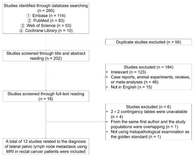
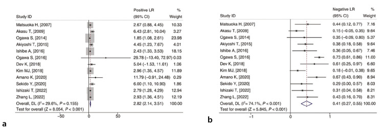
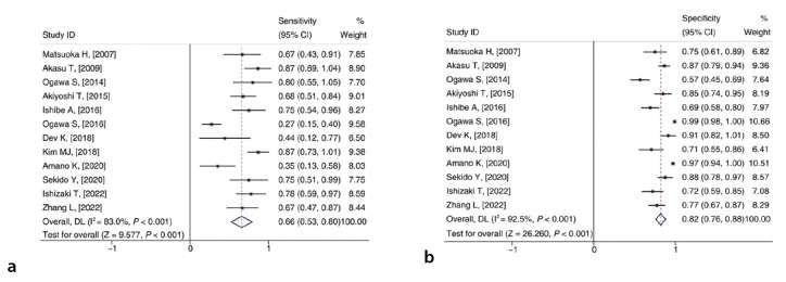
Results: This meta-analysis included 12 studies comprising 1,015 patients. The pooled sensitivity [95% confidence interval (CI)] and specificity (95% CI) of MRI for diagnosing LPLN metastasis were 0.66 (0.53, 0.80) and 0.82 (0.76, 0.88), respectively. The pooled positive likelihood ratio (LR) (95% CI) and negative LR (95% CI) were 2.82 (2.14, 3.51) and 0.41 (0.27, 0.55), respectively. The summary receiver operating characteristic curve indicated an area under the curve of 0.824. The quality of the included studies was acceptable according to the Quality Assessment of Diagnostic Accuracy Studies-2 tool. However, publication bias was present, as indicated by Deeks' funnel plot asymmetry test (P = 0.020). Considering that heterogeneity contributed to publication bias, a meta-regression analysis was conducted and revealed that heterogeneity could be influenced by sample size, with sample size negatively associated with sensitivity (coefficient: -0.002, P = 0.009) and positively associated with negative LR (coefficient: 0.002, P = 0.029).
Conclusion: Preoperative MRI demonstrates an acceptable ability to identify LPLN metastasis in patients with rectal carcinoma.
Clinical significance: Clinically, our findings support that preoperative MRI has acceptable diagnostic ability for LPLN metastasis in patients with rectal carcinoma. The preoperative application of MRI may aid in optimizing treatment strategies and improving prognosis in this population.
期刊介绍:
Diagnostic and Interventional Radiology (Diagn Interv Radiol) is the open access, online-only official publication of Turkish Society of Radiology. It is published bimonthly and the journal’s publication language is English.
The journal is a medium for original articles, reviews, pictorial essays, technical notes related to all fields of diagnostic and interventional radiology.

 求助内容:
求助内容: 应助结果提醒方式:


