{"title":"PVT1的敲低通过激活LATS2/Hippo信号通路抑制腔室和基底样乳腺癌亚型的细胞增殖。","authors":"Hai-Bo Zhang, Ying Zeng, Guo Wang","doi":"10.1186/s12957-025-03944-6","DOIUrl":null,"url":null,"abstract":"<p><strong>Background: </strong>Breast cancer (BC) is a malignant tumor seriously threatening women's health, while current approaches to BC treatment are challenged by the existence of drug resistance. Combination strategies of targeted therapy have been successfully applied in clinical BC treatment. However, whether there exist critical long non-coding RNAs (lncRNAs) responsible for BC pathogenesis and representing promising candidates for combined targeted therapy remains an issue.</p><p><strong>Methods: </strong>Public databases and bioinformatic methods were used to identify lncRNAs abnormally expressed among different subtypes of BC. The expression level of PVT1 was verified in collected clinical samples and representative cell lines. The role of PVT1 in BC cell proliferation was examined using MTS, plate clone formation, EdU and flow cytometry assay after small interfering RNA (siRNA) treatment. RNA sequencing was performed to investigate the potential molecular events regulated by PVT1. Western blot and immunofluorescence experiments were used to verify the activation of LATS2/Hippo signaling pathway after PVT1 knockdown. In addition, its activation was confirmed to mediate PVT1 function through rescue assay. The regulatory effect of PVT1 on LATS2 was investigated using mRNA stability experiments.</p><p><strong>Results: </strong>The expression level of PVT1 in BC tissues of luminal and basal-like subtypes was significantly higher than that in paracancerous tissues. PVT1 knockdown substantially inhibited the proliferation of BC cells in both subtypes. RNA sequencing revealed that Hippo signaling pathway might be the downstream target of PVT1. After PVT1 knockdown, both mRNA and protein levels of LATS2 were elevated which further decreased the distribution of YAP in cell nucleus, indicating the activation of Hippo signaling pathway. The proliferation inhibitory effect of PVT1 could be attenuated by simultaneous knockdown of LATS2. Furthermore, knockdown of PVT1 was demonstrated to significantly slow down the degradation rate of LATS2 mRNA.</p><p><strong>Conclusions: </strong>PVT1 level was significantly elevated in luminal and basal-like BC subtypes. Knockdown of PVT1 could inhibit cell proliferation of these two BC subtypes partly through activating LATS2/Hippo signaling pathway.</p>","PeriodicalId":23856,"journal":{"name":"World Journal of Surgical Oncology","volume":"23 1","pages":"289"},"PeriodicalIF":2.5000,"publicationDate":"2025-07-19","publicationTypes":"Journal Article","fieldsOfStudy":null,"isOpenAccess":false,"openAccessPdf":"https://www.ncbi.nlm.nih.gov/pmc/articles/PMC12275398/pdf/","citationCount":"0","resultStr":"{\"title\":\"Knockdown of PVT1 inhibits cell proliferation in luminal and basal-like breast cancer subtypes by activating LATS2/Hippo signaling pathway.\",\"authors\":\"Hai-Bo Zhang, Ying Zeng, Guo Wang\",\"doi\":\"10.1186/s12957-025-03944-6\",\"DOIUrl\":null,\"url\":null,\"abstract\":\"<p><strong>Background: </strong>Breast cancer (BC) is a malignant tumor seriously threatening women's health, while current approaches to BC treatment are challenged by the existence of drug resistance. Combination strategies of targeted therapy have been successfully applied in clinical BC treatment. However, whether there exist critical long non-coding RNAs (lncRNAs) responsible for BC pathogenesis and representing promising candidates for combined targeted therapy remains an issue.</p><p><strong>Methods: </strong>Public databases and bioinformatic methods were used to identify lncRNAs abnormally expressed among different subtypes of BC. The expression level of PVT1 was verified in collected clinical samples and representative cell lines. The role of PVT1 in BC cell proliferation was examined using MTS, plate clone formation, EdU and flow cytometry assay after small interfering RNA (siRNA) treatment. RNA sequencing was performed to investigate the potential molecular events regulated by PVT1. Western blot and immunofluorescence experiments were used to verify the activation of LATS2/Hippo signaling pathway after PVT1 knockdown. In addition, its activation was confirmed to mediate PVT1 function through rescue assay. The regulatory effect of PVT1 on LATS2 was investigated using mRNA stability experiments.</p><p><strong>Results: </strong>The expression level of PVT1 in BC tissues of luminal and basal-like subtypes was significantly higher than that in paracancerous tissues. PVT1 knockdown substantially inhibited the proliferation of BC cells in both subtypes. RNA sequencing revealed that Hippo signaling pathway might be the downstream target of PVT1. After PVT1 knockdown, both mRNA and protein levels of LATS2 were elevated which further decreased the distribution of YAP in cell nucleus, indicating the activation of Hippo signaling pathway. The proliferation inhibitory effect of PVT1 could be attenuated by simultaneous knockdown of LATS2. Furthermore, knockdown of PVT1 was demonstrated to significantly slow down the degradation rate of LATS2 mRNA.</p><p><strong>Conclusions: </strong>PVT1 level was significantly elevated in luminal and basal-like BC subtypes. Knockdown of PVT1 could inhibit cell proliferation of these two BC subtypes partly through activating LATS2/Hippo signaling pathway.</p>\",\"PeriodicalId\":23856,\"journal\":{\"name\":\"World Journal of Surgical Oncology\",\"volume\":\"23 1\",\"pages\":\"289\"},\"PeriodicalIF\":2.5000,\"publicationDate\":\"2025-07-19\",\"publicationTypes\":\"Journal Article\",\"fieldsOfStudy\":null,\"isOpenAccess\":false,\"openAccessPdf\":\"https://www.ncbi.nlm.nih.gov/pmc/articles/PMC12275398/pdf/\",\"citationCount\":\"0\",\"resultStr\":null,\"platform\":\"Semanticscholar\",\"paperid\":null,\"PeriodicalName\":\"World Journal of Surgical Oncology\",\"FirstCategoryId\":\"3\",\"ListUrlMain\":\"https://doi.org/10.1186/s12957-025-03944-6\",\"RegionNum\":3,\"RegionCategory\":\"医学\",\"ArticlePicture\":[],\"TitleCN\":null,\"AbstractTextCN\":null,\"PMCID\":null,\"EPubDate\":\"\",\"PubModel\":\"\",\"JCR\":\"Q3\",\"JCRName\":\"ONCOLOGY\",\"Score\":null,\"Total\":0}","platform":"Semanticscholar","paperid":null,"PeriodicalName":"World Journal of Surgical Oncology","FirstCategoryId":"3","ListUrlMain":"https://doi.org/10.1186/s12957-025-03944-6","RegionNum":3,"RegionCategory":"医学","ArticlePicture":[],"TitleCN":null,"AbstractTextCN":null,"PMCID":null,"EPubDate":"","PubModel":"","JCR":"Q3","JCRName":"ONCOLOGY","Score":null,"Total":0}
Knockdown of PVT1 inhibits cell proliferation in luminal and basal-like breast cancer subtypes by activating LATS2/Hippo signaling pathway.
Background: Breast cancer (BC) is a malignant tumor seriously threatening women's health, while current approaches to BC treatment are challenged by the existence of drug resistance. Combination strategies of targeted therapy have been successfully applied in clinical BC treatment. However, whether there exist critical long non-coding RNAs (lncRNAs) responsible for BC pathogenesis and representing promising candidates for combined targeted therapy remains an issue.
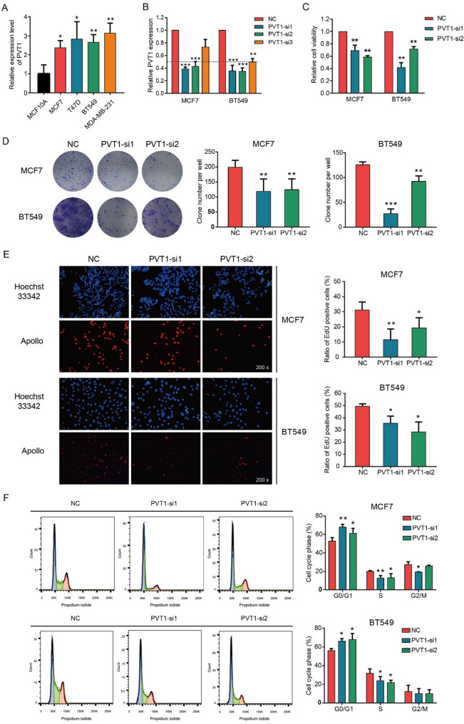
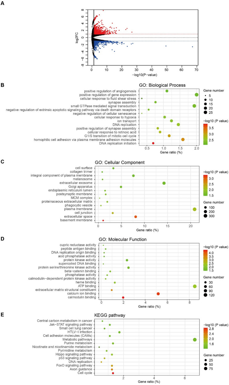
Methods: Public databases and bioinformatic methods were used to identify lncRNAs abnormally expressed among different subtypes of BC. The expression level of PVT1 was verified in collected clinical samples and representative cell lines. The role of PVT1 in BC cell proliferation was examined using MTS, plate clone formation, EdU and flow cytometry assay after small interfering RNA (siRNA) treatment. RNA sequencing was performed to investigate the potential molecular events regulated by PVT1. Western blot and immunofluorescence experiments were used to verify the activation of LATS2/Hippo signaling pathway after PVT1 knockdown. In addition, its activation was confirmed to mediate PVT1 function through rescue assay. The regulatory effect of PVT1 on LATS2 was investigated using mRNA stability experiments.
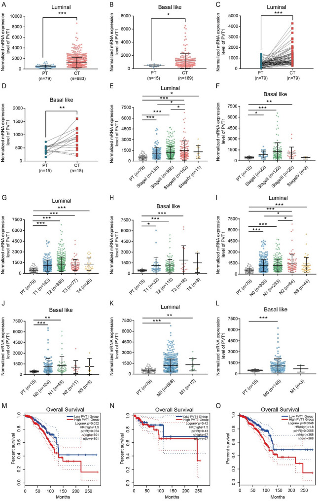
Results: The expression level of PVT1 in BC tissues of luminal and basal-like subtypes was significantly higher than that in paracancerous tissues. PVT1 knockdown substantially inhibited the proliferation of BC cells in both subtypes. RNA sequencing revealed that Hippo signaling pathway might be the downstream target of PVT1. After PVT1 knockdown, both mRNA and protein levels of LATS2 were elevated which further decreased the distribution of YAP in cell nucleus, indicating the activation of Hippo signaling pathway. The proliferation inhibitory effect of PVT1 could be attenuated by simultaneous knockdown of LATS2. Furthermore, knockdown of PVT1 was demonstrated to significantly slow down the degradation rate of LATS2 mRNA.
Conclusions: PVT1 level was significantly elevated in luminal and basal-like BC subtypes. Knockdown of PVT1 could inhibit cell proliferation of these two BC subtypes partly through activating LATS2/Hippo signaling pathway.
期刊介绍:
World Journal of Surgical Oncology publishes articles related to surgical oncology and its allied subjects, such as epidemiology, cancer research, biomarkers, prevention, pathology, radiology, cancer treatment, clinical trials, multimodality treatment and molecular biology. Emphasis is placed on original research articles. The journal also publishes significant clinical case reports, as well as balanced and timely reviews on selected topics.
Oncology is a multidisciplinary super-speciality of which surgical oncology forms an integral component, especially with solid tumors. Surgical oncologists around the world are involved in research extending from detecting the mechanisms underlying the causation of cancer, to its treatment and prevention. The role of a surgical oncologist extends across the whole continuum of care. With continued developments in diagnosis and treatment, the role of a surgical oncologist is ever-changing. Hence, World Journal of Surgical Oncology aims to keep readers abreast with latest developments that will ultimately influence the work of surgical oncologists.

 求助内容:
求助内容: 应助结果提醒方式:


