Delaney Loken, Luis F Goncalves, Mittun Patel, Nicholas Rubert
{"title":"使用超分辨率切片-体积重建增强胎儿MRI诊断食管闭锁。","authors":"Delaney Loken, Luis F Goncalves, Mittun Patel, Nicholas Rubert","doi":"10.1007/s00247-025-06309-z","DOIUrl":null,"url":null,"abstract":"<p><strong>Background: </strong>Prenatal diagnosis of esophageal atresia remains challenging, with indirect signs such as polyhydramnios, a small or absent stomach bubble, and a dilated upper esophageal pouch often inconsistently present. Only 10-40% of EA cases are diagnosed prenatally. Fetal MRI can overcome ultrasound limitations; however, constraints like motion can hinder evaluation of the esophagus.</p><p><strong>Methods: </strong>Super-resolution imaging with slice-to-volume reconstruction (SVR) is one approach that can improve image quality. This technique generates high-resolution 3D images from standard fetal MRI slices to enhance diagnostic accuracy.</p><p><strong>Aim: </strong>We present the application of super-resolution imaging with SVR to accurately diagnose EA and assess the presence or absence tracheoesophageal fistulas.</p><p><strong>Conclusion: </strong>This technique demonstrates significant potential for accurately delineating the relevant surgical anatomy, which can improve surgical planning.</p>","PeriodicalId":19755,"journal":{"name":"Pediatric Radiology","volume":" ","pages":"1943-1946"},"PeriodicalIF":2.3000,"publicationDate":"2025-08-01","publicationTypes":"Journal Article","fieldsOfStudy":null,"isOpenAccess":false,"openAccessPdf":"https://www.ncbi.nlm.nih.gov/pmc/articles/PMC12394271/pdf/","citationCount":"0","resultStr":"{\"title\":\"Enhanced fetal MRI diagnosis of esophageal atresia using super-resolution slice-to-volume reconstruction.\",\"authors\":\"Delaney Loken, Luis F Goncalves, Mittun Patel, Nicholas Rubert\",\"doi\":\"10.1007/s00247-025-06309-z\",\"DOIUrl\":null,\"url\":null,\"abstract\":\"<p><strong>Background: </strong>Prenatal diagnosis of esophageal atresia remains challenging, with indirect signs such as polyhydramnios, a small or absent stomach bubble, and a dilated upper esophageal pouch often inconsistently present. Only 10-40% of EA cases are diagnosed prenatally. Fetal MRI can overcome ultrasound limitations; however, constraints like motion can hinder evaluation of the esophagus.</p><p><strong>Methods: </strong>Super-resolution imaging with slice-to-volume reconstruction (SVR) is one approach that can improve image quality. This technique generates high-resolution 3D images from standard fetal MRI slices to enhance diagnostic accuracy.</p><p><strong>Aim: </strong>We present the application of super-resolution imaging with SVR to accurately diagnose EA and assess the presence or absence tracheoesophageal fistulas.</p><p><strong>Conclusion: </strong>This technique demonstrates significant potential for accurately delineating the relevant surgical anatomy, which can improve surgical planning.</p>\",\"PeriodicalId\":19755,\"journal\":{\"name\":\"Pediatric Radiology\",\"volume\":\" \",\"pages\":\"1943-1946\"},\"PeriodicalIF\":2.3000,\"publicationDate\":\"2025-08-01\",\"publicationTypes\":\"Journal Article\",\"fieldsOfStudy\":null,\"isOpenAccess\":false,\"openAccessPdf\":\"https://www.ncbi.nlm.nih.gov/pmc/articles/PMC12394271/pdf/\",\"citationCount\":\"0\",\"resultStr\":null,\"platform\":\"Semanticscholar\",\"paperid\":null,\"PeriodicalName\":\"Pediatric Radiology\",\"FirstCategoryId\":\"3\",\"ListUrlMain\":\"https://doi.org/10.1007/s00247-025-06309-z\",\"RegionNum\":3,\"RegionCategory\":\"医学\",\"ArticlePicture\":[],\"TitleCN\":null,\"AbstractTextCN\":null,\"PMCID\":null,\"EPubDate\":\"2025/7/19 0:00:00\",\"PubModel\":\"Epub\",\"JCR\":\"Q2\",\"JCRName\":\"PEDIATRICS\",\"Score\":null,\"Total\":0}","platform":"Semanticscholar","paperid":null,"PeriodicalName":"Pediatric Radiology","FirstCategoryId":"3","ListUrlMain":"https://doi.org/10.1007/s00247-025-06309-z","RegionNum":3,"RegionCategory":"医学","ArticlePicture":[],"TitleCN":null,"AbstractTextCN":null,"PMCID":null,"EPubDate":"2025/7/19 0:00:00","PubModel":"Epub","JCR":"Q2","JCRName":"PEDIATRICS","Score":null,"Total":0}
Enhanced fetal MRI diagnosis of esophageal atresia using super-resolution slice-to-volume reconstruction.
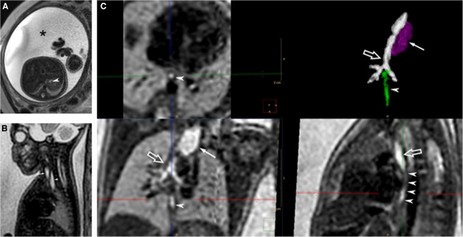
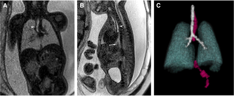
Background: Prenatal diagnosis of esophageal atresia remains challenging, with indirect signs such as polyhydramnios, a small or absent stomach bubble, and a dilated upper esophageal pouch often inconsistently present. Only 10-40% of EA cases are diagnosed prenatally. Fetal MRI can overcome ultrasound limitations; however, constraints like motion can hinder evaluation of the esophagus.
Methods: Super-resolution imaging with slice-to-volume reconstruction (SVR) is one approach that can improve image quality. This technique generates high-resolution 3D images from standard fetal MRI slices to enhance diagnostic accuracy.
Aim: We present the application of super-resolution imaging with SVR to accurately diagnose EA and assess the presence or absence tracheoesophageal fistulas.
Conclusion: This technique demonstrates significant potential for accurately delineating the relevant surgical anatomy, which can improve surgical planning.
期刊介绍:
Official Journal of the European Society of Pediatric Radiology, the Society for Pediatric Radiology and the Asian and Oceanic Society for Pediatric Radiology
Pediatric Radiology informs its readers of new findings and progress in all areas of pediatric imaging and in related fields. This is achieved by a blend of original papers, complemented by reviews that set out the present state of knowledge in a particular area of the specialty or summarize specific topics in which discussion has led to clear conclusions. Advances in technology, methodology, apparatus and auxiliary equipment are presented, and modifications of standard techniques are described.
Manuscripts submitted for publication must contain a statement to the effect that all human studies have been reviewed by the appropriate ethics committee and have therefore been performed in accordance with the ethical standards laid down in an appropriate version of the 1964 Declaration of Helsinki. It should also be stated clearly in the text that all persons gave their informed consent prior to their inclusion in the study. Details that might disclose the identity of the subjects under study should be omitted.

 求助内容:
求助内容: 应助结果提醒方式:


