Daniel Marchi Kieling, Amanda Hedel Koerich, Romildo Antonio Dos Santos Júnior, Paulo Moacir Mesquita Filho
{"title":"松果体囊肿合并中风和脑积水1例报告。","authors":"Daniel Marchi Kieling, Amanda Hedel Koerich, Romildo Antonio Dos Santos Júnior, Paulo Moacir Mesquita Filho","doi":"10.12865/CHSJ.51.01.19","DOIUrl":null,"url":null,"abstract":"<p><p>Pineal cysts (PCs) are common findings on (Magnetic resonance Imaging) MRI, often incidental in females and asymptomatic throughout life. Rare complications, like pineal apoplexy with acute hydrocephalus, require differential diagnosis and urgent intervention. We report a 19-year-old male with a progressive headache and visual decline. MRI showed a 2.3 cm pineal cyst with hemorrhagic features (apoplexy) causing hydrocephalus. He underwent endoscopic third ventriculostomy and microsurgical resection, both successful. Postoperatively, symptoms resolved completely. Though often benign, complicated PCs can be life-threatening. This case highlights their management, aiding understanding of etiologies, differential diagnoses, and treatments, enhancing medical knowledge.</p>","PeriodicalId":93963,"journal":{"name":"Current health sciences journal","volume":"51 1","pages":"164-169"},"PeriodicalIF":0.0000,"publicationDate":"2025-01-01","publicationTypes":"Journal Article","fieldsOfStudy":null,"isOpenAccess":false,"openAccessPdf":"https://www.ncbi.nlm.nih.gov/pmc/articles/PMC12264991/pdf/","citationCount":"0","resultStr":"{\"title\":\"Pineal Cyst Associated with Apoplexy and Hydrocephalus: A Case Report.\",\"authors\":\"Daniel Marchi Kieling, Amanda Hedel Koerich, Romildo Antonio Dos Santos Júnior, Paulo Moacir Mesquita Filho\",\"doi\":\"10.12865/CHSJ.51.01.19\",\"DOIUrl\":null,\"url\":null,\"abstract\":\"<p><p>Pineal cysts (PCs) are common findings on (Magnetic resonance Imaging) MRI, often incidental in females and asymptomatic throughout life. Rare complications, like pineal apoplexy with acute hydrocephalus, require differential diagnosis and urgent intervention. We report a 19-year-old male with a progressive headache and visual decline. MRI showed a 2.3 cm pineal cyst with hemorrhagic features (apoplexy) causing hydrocephalus. He underwent endoscopic third ventriculostomy and microsurgical resection, both successful. Postoperatively, symptoms resolved completely. Though often benign, complicated PCs can be life-threatening. This case highlights their management, aiding understanding of etiologies, differential diagnoses, and treatments, enhancing medical knowledge.</p>\",\"PeriodicalId\":93963,\"journal\":{\"name\":\"Current health sciences journal\",\"volume\":\"51 1\",\"pages\":\"164-169\"},\"PeriodicalIF\":0.0000,\"publicationDate\":\"2025-01-01\",\"publicationTypes\":\"Journal Article\",\"fieldsOfStudy\":null,\"isOpenAccess\":false,\"openAccessPdf\":\"https://www.ncbi.nlm.nih.gov/pmc/articles/PMC12264991/pdf/\",\"citationCount\":\"0\",\"resultStr\":null,\"platform\":\"Semanticscholar\",\"paperid\":null,\"PeriodicalName\":\"Current health sciences journal\",\"FirstCategoryId\":\"1085\",\"ListUrlMain\":\"https://doi.org/10.12865/CHSJ.51.01.19\",\"RegionNum\":0,\"RegionCategory\":null,\"ArticlePicture\":[],\"TitleCN\":null,\"AbstractTextCN\":null,\"PMCID\":null,\"EPubDate\":\"2025/3/31 0:00:00\",\"PubModel\":\"Epub\",\"JCR\":\"\",\"JCRName\":\"\",\"Score\":null,\"Total\":0}","platform":"Semanticscholar","paperid":null,"PeriodicalName":"Current health sciences journal","FirstCategoryId":"1085","ListUrlMain":"https://doi.org/10.12865/CHSJ.51.01.19","RegionNum":0,"RegionCategory":null,"ArticlePicture":[],"TitleCN":null,"AbstractTextCN":null,"PMCID":null,"EPubDate":"2025/3/31 0:00:00","PubModel":"Epub","JCR":"","JCRName":"","Score":null,"Total":0}
Pineal Cyst Associated with Apoplexy and Hydrocephalus: A Case Report.
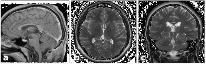
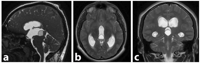
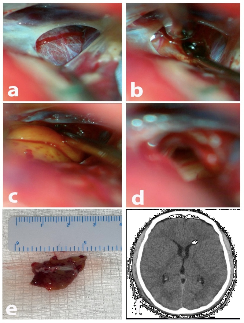
Pineal cysts (PCs) are common findings on (Magnetic resonance Imaging) MRI, often incidental in females and asymptomatic throughout life. Rare complications, like pineal apoplexy with acute hydrocephalus, require differential diagnosis and urgent intervention. We report a 19-year-old male with a progressive headache and visual decline. MRI showed a 2.3 cm pineal cyst with hemorrhagic features (apoplexy) causing hydrocephalus. He underwent endoscopic third ventriculostomy and microsurgical resection, both successful. Postoperatively, symptoms resolved completely. Though often benign, complicated PCs can be life-threatening. This case highlights their management, aiding understanding of etiologies, differential diagnoses, and treatments, enhancing medical knowledge.

 求助内容:
求助内容: 应助结果提醒方式:


