Boaz Karmazyn, Matthew M Jones, Lisa R Delaney, Ralph A Hicks, Ann E Freshour, Megan B Marine, Shannon L Thompson, S Gregory Jennings, George J Eckert, Monica M Forbes-Amrhein
{"title":"2岁以下儿童肋软骨连接处的变异。","authors":"Boaz Karmazyn, Matthew M Jones, Lisa R Delaney, Ralph A Hicks, Ann E Freshour, Megan B Marine, Shannon L Thompson, S Gregory Jennings, George J Eckert, Monica M Forbes-Amrhein","doi":"10.1007/s00247-025-06316-0","DOIUrl":null,"url":null,"abstract":"<p><strong>Background: </strong>Costochondral junction fractures are considered specific for child abuse and typically heal with a deformed costochondral junction.</p><p><strong>Objective: </strong>To evaluate the types, location, and incidence of costochondral junction variations that can mimic fractures.</p><p><strong>Materials and methods: </strong>A 15-year retrospective study was conducted on children under 2 years of age who underwent chest and abdominal computerized tomography (CT) scans for pneumonia, fever, congenital lung disease, pain, or appendicitis. Randomized selection included 120 chest and 120 abdominal CT scans. Demographic and clinical information was obtained from medical record reviews. Two pediatric radiologists independently reviewed the studies and indicated the presence and location of costochondral junction variation patterns (spurs), and fissure, horizontal lucency, corner, or bucket handle as identified on two consecutive slices on axial views. Disagreements were resolved by a third radiologist. We excluded patients with underlying medical conditions that could affect the skeleton and studies with motion artifacts. A t-test was used to evaluate the relationships between age, CT slice thickness, and the diagnosis of costochondral junction variations. Kappa statistics were used to evaluate agreement.</p><p><strong>Results: </strong>A total of 123 children were excluded due to motion artifacts (n = 30), trauma (n = 31), being evaluated for child abuse (n = 3), slice thickness of 5 mm (n = 1), and underlying medical conditions (n = 58). The final group included 117 children (73 males and 44 females) with an average age of 1 year; 64 had chest and 53 abdominal CT scans. Agreement was fair (kappa = 0.29) at the patient level and poor at the rib level (kappa = 0-0.64). The final number of variations, after resolving disagreements with a third radiologist, was 46 of costochondral junction variations in 19 children (16.2%, 19/117); all were costochondral junction spurs at the levels of the second to eighth ribs. Costochondral junction variations were significantly more common in younger children (average 0.7 ± 0.6 years vs. 1.1 ± 0.6 years, P = 0.024) and when there was thinner CT slice thickness (average 1.6 ± 1.4 mm vs. 2.5 ± 1.5 mm, P = 0.041).</p><p><strong>Conclusion: </strong>Costochondral junction variations were identified in 16.2% of children under 2 years of age, and some may mimic healing costochondral junction fractures. There was only fair agreement between radiologists.</p>","PeriodicalId":19755,"journal":{"name":"Pediatric Radiology","volume":" ","pages":"1883-1890"},"PeriodicalIF":2.3000,"publicationDate":"2025-08-01","publicationTypes":"Journal Article","fieldsOfStudy":null,"isOpenAccess":false,"openAccessPdf":"https://www.ncbi.nlm.nih.gov/pmc/articles/PMC12394368/pdf/","citationCount":"0","resultStr":"{\"title\":\"Costochondral junction variations in children younger than 2 years.\",\"authors\":\"Boaz Karmazyn, Matthew M Jones, Lisa R Delaney, Ralph A Hicks, Ann E Freshour, Megan B Marine, Shannon L Thompson, S Gregory Jennings, George J Eckert, Monica M Forbes-Amrhein\",\"doi\":\"10.1007/s00247-025-06316-0\",\"DOIUrl\":null,\"url\":null,\"abstract\":\"<p><strong>Background: </strong>Costochondral junction fractures are considered specific for child abuse and typically heal with a deformed costochondral junction.</p><p><strong>Objective: </strong>To evaluate the types, location, and incidence of costochondral junction variations that can mimic fractures.</p><p><strong>Materials and methods: </strong>A 15-year retrospective study was conducted on children under 2 years of age who underwent chest and abdominal computerized tomography (CT) scans for pneumonia, fever, congenital lung disease, pain, or appendicitis. Randomized selection included 120 chest and 120 abdominal CT scans. Demographic and clinical information was obtained from medical record reviews. Two pediatric radiologists independently reviewed the studies and indicated the presence and location of costochondral junction variation patterns (spurs), and fissure, horizontal lucency, corner, or bucket handle as identified on two consecutive slices on axial views. Disagreements were resolved by a third radiologist. We excluded patients with underlying medical conditions that could affect the skeleton and studies with motion artifacts. A t-test was used to evaluate the relationships between age, CT slice thickness, and the diagnosis of costochondral junction variations. Kappa statistics were used to evaluate agreement.</p><p><strong>Results: </strong>A total of 123 children were excluded due to motion artifacts (n = 30), trauma (n = 31), being evaluated for child abuse (n = 3), slice thickness of 5 mm (n = 1), and underlying medical conditions (n = 58). The final group included 117 children (73 males and 44 females) with an average age of 1 year; 64 had chest and 53 abdominal CT scans. Agreement was fair (kappa = 0.29) at the patient level and poor at the rib level (kappa = 0-0.64). The final number of variations, after resolving disagreements with a third radiologist, was 46 of costochondral junction variations in 19 children (16.2%, 19/117); all were costochondral junction spurs at the levels of the second to eighth ribs. Costochondral junction variations were significantly more common in younger children (average 0.7 ± 0.6 years vs. 1.1 ± 0.6 years, P = 0.024) and when there was thinner CT slice thickness (average 1.6 ± 1.4 mm vs. 2.5 ± 1.5 mm, P = 0.041).</p><p><strong>Conclusion: </strong>Costochondral junction variations were identified in 16.2% of children under 2 years of age, and some may mimic healing costochondral junction fractures. There was only fair agreement between radiologists.</p>\",\"PeriodicalId\":19755,\"journal\":{\"name\":\"Pediatric Radiology\",\"volume\":\" \",\"pages\":\"1883-1890\"},\"PeriodicalIF\":2.3000,\"publicationDate\":\"2025-08-01\",\"publicationTypes\":\"Journal Article\",\"fieldsOfStudy\":null,\"isOpenAccess\":false,\"openAccessPdf\":\"https://www.ncbi.nlm.nih.gov/pmc/articles/PMC12394368/pdf/\",\"citationCount\":\"0\",\"resultStr\":null,\"platform\":\"Semanticscholar\",\"paperid\":null,\"PeriodicalName\":\"Pediatric Radiology\",\"FirstCategoryId\":\"3\",\"ListUrlMain\":\"https://doi.org/10.1007/s00247-025-06316-0\",\"RegionNum\":3,\"RegionCategory\":\"医学\",\"ArticlePicture\":[],\"TitleCN\":null,\"AbstractTextCN\":null,\"PMCID\":null,\"EPubDate\":\"2025/7/17 0:00:00\",\"PubModel\":\"Epub\",\"JCR\":\"Q2\",\"JCRName\":\"PEDIATRICS\",\"Score\":null,\"Total\":0}","platform":"Semanticscholar","paperid":null,"PeriodicalName":"Pediatric Radiology","FirstCategoryId":"3","ListUrlMain":"https://doi.org/10.1007/s00247-025-06316-0","RegionNum":3,"RegionCategory":"医学","ArticlePicture":[],"TitleCN":null,"AbstractTextCN":null,"PMCID":null,"EPubDate":"2025/7/17 0:00:00","PubModel":"Epub","JCR":"Q2","JCRName":"PEDIATRICS","Score":null,"Total":0}
引用次数: 0
摘要
背景:肋软骨连接处骨折被认为是儿童虐待特有的,通常以畸形的肋软骨连接处愈合。目的:探讨可模拟骨折的肋软骨连接处变异的类型、位置和发生率。材料和方法:对2岁以下儿童进行了为期15年的回顾性研究,这些儿童因肺炎、发热、先天性肺病、疼痛或阑尾炎进行了胸部和腹部计算机断层扫描(CT)。随机选择120个胸部和120个腹部CT扫描。从医疗记录审查中获得人口统计和临床信息。两名儿科放射科医生独立审查了这些研究,并指出肋软骨连接处变异模式(骨刺)、裂缝、水平透光、角状或桶状柄的存在和位置在连续两张轴位片上被识别出来。分歧由第三位放射科医生解决。我们排除了可能影响骨骼的潜在疾病患者和运动伪影研究。采用t检验评价年龄、CT层厚度与肋软骨连接处病变诊断之间的关系。采用Kappa统计来评价一致性。结果:共有123名儿童因运动假影(n = 30)、创伤(n = 31)、儿童虐待评估(n = 3)、切片厚度为5mm (n = 1)和潜在医疗条件(n = 58)而被排除。最后一组117名儿童(男73名,女44名),平均年龄1岁;64例胸部CT, 53例腹部CT。在患者水平上,一致性是一般的(kappa = 0.29),在肋骨水平上一致性较差(kappa = 0-0.64)。在与第三位放射科医生解决分歧后,最终变异数为19例儿童中46例肋软骨结变异(16.2%,19/117);在第二至第八肋骨的水平均为肋软骨结合部刺。肋软骨结变异在年龄较小的儿童(平均0.7±0.6岁vs 1.1±0.6岁,P = 0.024)和CT层厚度较薄的儿童(平均1.6±1.4 mm vs 2.5±1.5 mm, P = 0.041)中更为常见。结论:16.2%的2岁以下儿童存在肋软骨连接处变异,其中一些可能与愈合性肋软骨连接处骨折相似。放射科医生之间只有公平的共识。
Costochondral junction variations in children younger than 2 years.
Background: Costochondral junction fractures are considered specific for child abuse and typically heal with a deformed costochondral junction.
Objective: To evaluate the types, location, and incidence of costochondral junction variations that can mimic fractures.
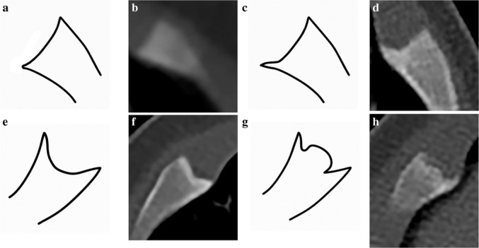
Materials and methods: A 15-year retrospective study was conducted on children under 2 years of age who underwent chest and abdominal computerized tomography (CT) scans for pneumonia, fever, congenital lung disease, pain, or appendicitis. Randomized selection included 120 chest and 120 abdominal CT scans. Demographic and clinical information was obtained from medical record reviews. Two pediatric radiologists independently reviewed the studies and indicated the presence and location of costochondral junction variation patterns (spurs), and fissure, horizontal lucency, corner, or bucket handle as identified on two consecutive slices on axial views. Disagreements were resolved by a third radiologist. We excluded patients with underlying medical conditions that could affect the skeleton and studies with motion artifacts. A t-test was used to evaluate the relationships between age, CT slice thickness, and the diagnosis of costochondral junction variations. Kappa statistics were used to evaluate agreement.
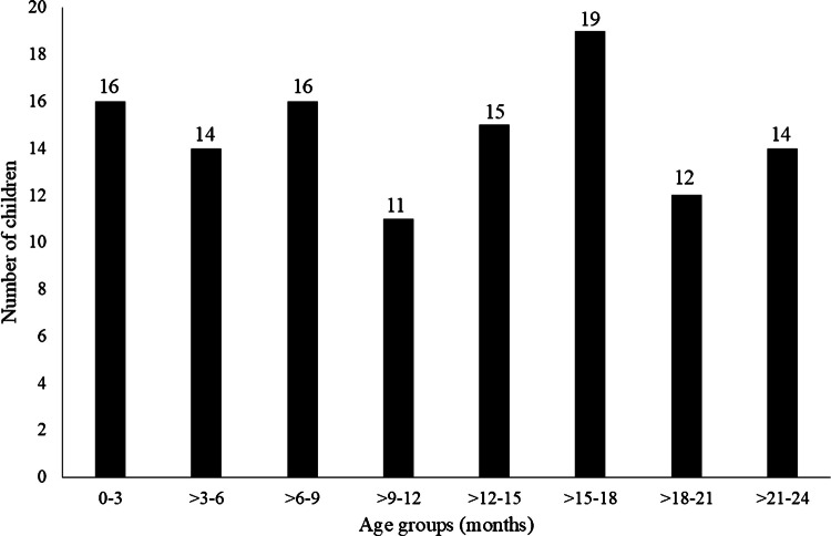
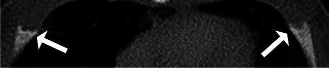
Results: A total of 123 children were excluded due to motion artifacts (n = 30), trauma (n = 31), being evaluated for child abuse (n = 3), slice thickness of 5 mm (n = 1), and underlying medical conditions (n = 58). The final group included 117 children (73 males and 44 females) with an average age of 1 year; 64 had chest and 53 abdominal CT scans. Agreement was fair (kappa = 0.29) at the patient level and poor at the rib level (kappa = 0-0.64). The final number of variations, after resolving disagreements with a third radiologist, was 46 of costochondral junction variations in 19 children (16.2%, 19/117); all were costochondral junction spurs at the levels of the second to eighth ribs. Costochondral junction variations were significantly more common in younger children (average 0.7 ± 0.6 years vs. 1.1 ± 0.6 years, P = 0.024) and when there was thinner CT slice thickness (average 1.6 ± 1.4 mm vs. 2.5 ± 1.5 mm, P = 0.041).
Conclusion: Costochondral junction variations were identified in 16.2% of children under 2 years of age, and some may mimic healing costochondral junction fractures. There was only fair agreement between radiologists.
期刊介绍:
Official Journal of the European Society of Pediatric Radiology, the Society for Pediatric Radiology and the Asian and Oceanic Society for Pediatric Radiology
Pediatric Radiology informs its readers of new findings and progress in all areas of pediatric imaging and in related fields. This is achieved by a blend of original papers, complemented by reviews that set out the present state of knowledge in a particular area of the specialty or summarize specific topics in which discussion has led to clear conclusions. Advances in technology, methodology, apparatus and auxiliary equipment are presented, and modifications of standard techniques are described.
Manuscripts submitted for publication must contain a statement to the effect that all human studies have been reviewed by the appropriate ethics committee and have therefore been performed in accordance with the ethical standards laid down in an appropriate version of the 1964 Declaration of Helsinki. It should also be stated clearly in the text that all persons gave their informed consent prior to their inclusion in the study. Details that might disclose the identity of the subjects under study should be omitted.

 求助内容:
求助内容: 应助结果提醒方式:


