Sara Ahmadi Badi, Hananeh Tavakoli Aval, Hamid Reza Moradi, Amin Malek, Seyed Amirhesam Seyedi, Mehdi Davari, Ahmad Bereimipour, Soghra Khani, Shohreh Khatami, Seyed Davar Siadat
{"title":"嗜黏液阿克曼氏菌和prausnitzfaecalibacterium通过靶向hepcidin -铁转运蛋白轴对CCl 4诱导肝纤维化小鼠肝损伤的活清和无细胞上清干预的比较研究","authors":"Sara Ahmadi Badi, Hananeh Tavakoli Aval, Hamid Reza Moradi, Amin Malek, Seyed Amirhesam Seyedi, Mehdi Davari, Ahmad Bereimipour, Soghra Khani, Shohreh Khatami, Seyed Davar Siadat","doi":"10.1186/s13099-025-00728-x","DOIUrl":null,"url":null,"abstract":"<p><strong>Background: </strong>liver fibrosis is associated with dysregulated iron homeostasis regulated by the hepcidin-ferroportin axis, and dysbiotic gut microbiota. This study aimed to investigate the preventive and ameliorative effects of live and cell-free supernatant (CFS) forms of Akkermansia muciniphila and Faecalibacterium prausnitzii, as important gut microbiota members, on liver fibrosis by targeting the hepcidin-ferroportin axis in both in vitro and in vivo models.</p><p><strong>Methods: </strong>At the in vitro level, the effects of A. muciniphila and F. prausnitzii on the expression of collagen type I alpha 1 (COL1A1) and ferroportin (SLC40A1) transcripts in hepatic stellate cells (HSCs) were evaluated in transforming growth factor beta (TGFβ)-activated LX-2 cells, a human hepatic stellate cell line. In vivo, male C57BL/6 mice were intraperitoneally (IP) injected with 10% carbon tetrachloride (CCl₄) twice weekly for 6 weeks to establish the liver fibrosis model. Administration of live and CFS forms of A. muciniphila and F. prausnitzii was initiated 10 days before CCl₄ injection and continued until the end of the experiment. Liver injury and fibrosis were assessed using serum markers, hematoxylin and eosin (H&E), and Masson's trichrome staining. Reverse transcription-quantitative polymerase chain reaction (RT-qPCR) and immunohistochemistry (IHC) were used to evaluate the effects of the interventions on gene expression related to the hepcidin-ferroportin axis in liver, colon and brain samples. Additionally, qPCR was used to determine alterations in the relative abundance of key gut microbiota members in fecal samples.</p><p><strong>Results: </strong>Both A. muciniphila and F. prausnitzii, as well as their CFS, significantly downregulated COL1A1 expression in TGFβ-activated LX-2 cells, accompanied by reduced alpha-smooth muscle actin (α-SMA) protein expression in liver tissue. In vivo, intervention with F. prausnitzii, particularly its CFS, led to a greater induction of hepatic hepcidin and ferroportin expression compared to A. muciniphila and its CFS. Serum liver injury markers (alanine aminotransferase (ALT), aspartate aminotransferase (AST), lactate dehydrogenase (LDH)) and iron levels were markedly improved following treatment with live F. prausnitzii and its CFS. Additionally, F. prausnitzii CFS significantly enhanced hepcidin gene expression in brain tissue, suggesting broader systemic benefits.</p><p><strong>Conclusion: </strong>We demonstrated that F. prausnitzii and its CFS had greater beneficial potential than A. muciniphila and its CFS in the prevention and amelioration of liver fibrosis, likely through modulation of the hepcidin-ferroportin axis. These findings may support the development of next-generation probiotics and postbiotics for liver injury, which warrants further investigation.</p>","PeriodicalId":12833,"journal":{"name":"Gut Pathogens","volume":"17 1","pages":"54"},"PeriodicalIF":4.0000,"publicationDate":"2025-07-17","publicationTypes":"Journal Article","fieldsOfStudy":null,"isOpenAccess":false,"openAccessPdf":"https://www.ncbi.nlm.nih.gov/pmc/articles/PMC12273000/pdf/","citationCount":"0","resultStr":"{\"title\":\"Comparative study of liver injury protection by Akkermansia muciniphila and Faecalibacterium prausnitzii interventions in live and cell-free supernatant forms via targeting the hepcidin - ferroportin axis in mice with CCl₄-induced liver fibrosis.\",\"authors\":\"Sara Ahmadi Badi, Hananeh Tavakoli Aval, Hamid Reza Moradi, Amin Malek, Seyed Amirhesam Seyedi, Mehdi Davari, Ahmad Bereimipour, Soghra Khani, Shohreh Khatami, Seyed Davar Siadat\",\"doi\":\"10.1186/s13099-025-00728-x\",\"DOIUrl\":null,\"url\":null,\"abstract\":\"<p><strong>Background: </strong>liver fibrosis is associated with dysregulated iron homeostasis regulated by the hepcidin-ferroportin axis, and dysbiotic gut microbiota. This study aimed to investigate the preventive and ameliorative effects of live and cell-free supernatant (CFS) forms of Akkermansia muciniphila and Faecalibacterium prausnitzii, as important gut microbiota members, on liver fibrosis by targeting the hepcidin-ferroportin axis in both in vitro and in vivo models.</p><p><strong>Methods: </strong>At the in vitro level, the effects of A. muciniphila and F. prausnitzii on the expression of collagen type I alpha 1 (COL1A1) and ferroportin (SLC40A1) transcripts in hepatic stellate cells (HSCs) were evaluated in transforming growth factor beta (TGFβ)-activated LX-2 cells, a human hepatic stellate cell line. In vivo, male C57BL/6 mice were intraperitoneally (IP) injected with 10% carbon tetrachloride (CCl₄) twice weekly for 6 weeks to establish the liver fibrosis model. Administration of live and CFS forms of A. muciniphila and F. prausnitzii was initiated 10 days before CCl₄ injection and continued until the end of the experiment. Liver injury and fibrosis were assessed using serum markers, hematoxylin and eosin (H&E), and Masson's trichrome staining. Reverse transcription-quantitative polymerase chain reaction (RT-qPCR) and immunohistochemistry (IHC) were used to evaluate the effects of the interventions on gene expression related to the hepcidin-ferroportin axis in liver, colon and brain samples. Additionally, qPCR was used to determine alterations in the relative abundance of key gut microbiota members in fecal samples.</p><p><strong>Results: </strong>Both A. muciniphila and F. prausnitzii, as well as their CFS, significantly downregulated COL1A1 expression in TGFβ-activated LX-2 cells, accompanied by reduced alpha-smooth muscle actin (α-SMA) protein expression in liver tissue. In vivo, intervention with F. prausnitzii, particularly its CFS, led to a greater induction of hepatic hepcidin and ferroportin expression compared to A. muciniphila and its CFS. Serum liver injury markers (alanine aminotransferase (ALT), aspartate aminotransferase (AST), lactate dehydrogenase (LDH)) and iron levels were markedly improved following treatment with live F. prausnitzii and its CFS. Additionally, F. prausnitzii CFS significantly enhanced hepcidin gene expression in brain tissue, suggesting broader systemic benefits.</p><p><strong>Conclusion: </strong>We demonstrated that F. prausnitzii and its CFS had greater beneficial potential than A. muciniphila and its CFS in the prevention and amelioration of liver fibrosis, likely through modulation of the hepcidin-ferroportin axis. These findings may support the development of next-generation probiotics and postbiotics for liver injury, which warrants further investigation.</p>\",\"PeriodicalId\":12833,\"journal\":{\"name\":\"Gut Pathogens\",\"volume\":\"17 1\",\"pages\":\"54\"},\"PeriodicalIF\":4.0000,\"publicationDate\":\"2025-07-17\",\"publicationTypes\":\"Journal Article\",\"fieldsOfStudy\":null,\"isOpenAccess\":false,\"openAccessPdf\":\"https://www.ncbi.nlm.nih.gov/pmc/articles/PMC12273000/pdf/\",\"citationCount\":\"0\",\"resultStr\":null,\"platform\":\"Semanticscholar\",\"paperid\":null,\"PeriodicalName\":\"Gut Pathogens\",\"FirstCategoryId\":\"3\",\"ListUrlMain\":\"https://doi.org/10.1186/s13099-025-00728-x\",\"RegionNum\":3,\"RegionCategory\":\"医学\",\"ArticlePicture\":[],\"TitleCN\":null,\"AbstractTextCN\":null,\"PMCID\":null,\"EPubDate\":\"\",\"PubModel\":\"\",\"JCR\":\"Q1\",\"JCRName\":\"GASTROENTEROLOGY & HEPATOLOGY\",\"Score\":null,\"Total\":0}","platform":"Semanticscholar","paperid":null,"PeriodicalName":"Gut Pathogens","FirstCategoryId":"3","ListUrlMain":"https://doi.org/10.1186/s13099-025-00728-x","RegionNum":3,"RegionCategory":"医学","ArticlePicture":[],"TitleCN":null,"AbstractTextCN":null,"PMCID":null,"EPubDate":"","PubModel":"","JCR":"Q1","JCRName":"GASTROENTEROLOGY & HEPATOLOGY","Score":null,"Total":0}
Comparative study of liver injury protection by Akkermansia muciniphila and Faecalibacterium prausnitzii interventions in live and cell-free supernatant forms via targeting the hepcidin - ferroportin axis in mice with CCl₄-induced liver fibrosis.
Background: liver fibrosis is associated with dysregulated iron homeostasis regulated by the hepcidin-ferroportin axis, and dysbiotic gut microbiota. This study aimed to investigate the preventive and ameliorative effects of live and cell-free supernatant (CFS) forms of Akkermansia muciniphila and Faecalibacterium prausnitzii, as important gut microbiota members, on liver fibrosis by targeting the hepcidin-ferroportin axis in both in vitro and in vivo models.
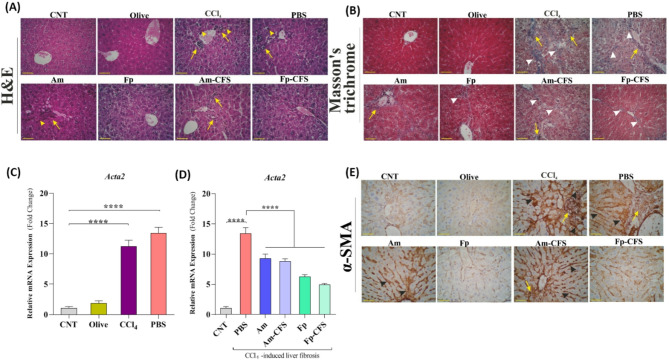
Methods: At the in vitro level, the effects of A. muciniphila and F. prausnitzii on the expression of collagen type I alpha 1 (COL1A1) and ferroportin (SLC40A1) transcripts in hepatic stellate cells (HSCs) were evaluated in transforming growth factor beta (TGFβ)-activated LX-2 cells, a human hepatic stellate cell line. In vivo, male C57BL/6 mice were intraperitoneally (IP) injected with 10% carbon tetrachloride (CCl₄) twice weekly for 6 weeks to establish the liver fibrosis model. Administration of live and CFS forms of A. muciniphila and F. prausnitzii was initiated 10 days before CCl₄ injection and continued until the end of the experiment. Liver injury and fibrosis were assessed using serum markers, hematoxylin and eosin (H&E), and Masson's trichrome staining. Reverse transcription-quantitative polymerase chain reaction (RT-qPCR) and immunohistochemistry (IHC) were used to evaluate the effects of the interventions on gene expression related to the hepcidin-ferroportin axis in liver, colon and brain samples. Additionally, qPCR was used to determine alterations in the relative abundance of key gut microbiota members in fecal samples.
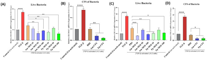
Results: Both A. muciniphila and F. prausnitzii, as well as their CFS, significantly downregulated COL1A1 expression in TGFβ-activated LX-2 cells, accompanied by reduced alpha-smooth muscle actin (α-SMA) protein expression in liver tissue. In vivo, intervention with F. prausnitzii, particularly its CFS, led to a greater induction of hepatic hepcidin and ferroportin expression compared to A. muciniphila and its CFS. Serum liver injury markers (alanine aminotransferase (ALT), aspartate aminotransferase (AST), lactate dehydrogenase (LDH)) and iron levels were markedly improved following treatment with live F. prausnitzii and its CFS. Additionally, F. prausnitzii CFS significantly enhanced hepcidin gene expression in brain tissue, suggesting broader systemic benefits.
Conclusion: We demonstrated that F. prausnitzii and its CFS had greater beneficial potential than A. muciniphila and its CFS in the prevention and amelioration of liver fibrosis, likely through modulation of the hepcidin-ferroportin axis. These findings may support the development of next-generation probiotics and postbiotics for liver injury, which warrants further investigation.
Gut PathogensGASTROENTEROLOGY & HEPATOLOGY-MICROBIOLOGY
CiteScore
7.70
自引率
2.40%
发文量
43
期刊介绍:
Gut Pathogens is a fast publishing, inclusive and prominent international journal which recognizes the need for a publishing platform uniquely tailored to reflect the full breadth of research in the biology and medicine of pathogens, commensals and functional microbiota of the gut. The journal publishes basic, clinical and cutting-edge research on all aspects of the above mentioned organisms including probiotic bacteria and yeasts and their products. The scope also covers the related ecology, molecular genetics, physiology and epidemiology of these microbes. The journal actively invites timely reports on the novel aspects of genomics, metagenomics, microbiota profiling and systems biology.
Gut Pathogens will also consider, at the discretion of the editors, descriptive studies identifying a new genome sequence of a gut microbe or a series of related microbes (such as those obtained from new hosts, niches, settings, outbreaks and epidemics) and those obtained from single or multiple hosts at one or different time points (chronological evolution).
 求助内容:
求助内容: 应助结果提醒方式:


