两种纳米复合树脂对人牙龈成纤维细胞的生物学效应(体外研究)
Q1 Medicine
Journal of oral biology and craniofacial research
Pub Date : 2025-07-16
DOI:10.1016/j.jobcr.2025.07.006
引用次数: 0
摘要
目的研究两种不同纳米复合树脂对人原代牙龈成纤维细胞的遗传毒性和细胞毒性。方法对hgf进行分离鉴定,并将其分为3组。对照组由未经处理的hgf、OMN组和3M Filtek Z350 xt提取物处理的hgf (ESPE组)组成。在HGF预孵育72 h和24 h后,观察细胞活力、细胞死亡方式及白细胞介素-1β (IL-1β)和白细胞介素-6 (IL-6)的表达。结果分离的hgf具有一定的特征。在大多数浓度下,OMN组的HGF活力显著高于ESPE组。与对照组相比,ESPE组和OMN组在72 h时的总细胞死亡率更高,ESPE组比OMN组更高。此外,OMN组和ESPE组在72 h时IL-1β和IL-6水平均高于对照组。结论ESPE对HGF的细胞毒和基因毒作用比OMN更显著。本文章由计算机程序翻译,如有差异,请以英文原文为准。
Biological effects of two nano-composite resins on human gingival fibroblast (an in vitro study)
Objective
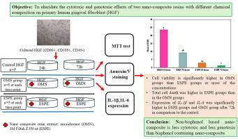
To elucidate the genotoxic and cytotoxic effects of two different nano-composite resins on primary human gingival fibroblasts (HGFs).
Methods
The HGFs were isolated, characterized, and classified into 3 groups. The control group consisted of untreated HGFs, the Omnichroma extract treated HGFs (the OMN group), and the 3M Filtek Z350 xt extract treated HGFs (ESPE group). The cell viability, mode of cell death and expression of interleukin-1β (IL-1β) and interleukin-6 (IL-6) were investigated after 72 h and 24 h of resin extracts' pre-incubation with HGF.
Results
The isolated HGFs were characterized. The HGF viability was significantly higher in the OMN groups than ESPE groups at most of the concentrations. Total cell death was higher in the ESPE groups and at the OMN group at 72 h in comparison to the control and was higher in the ESPE groups compared to OMN groups. Furthermore, the IL-1β and IL-6 levels in the OMN group at 72 h and in the ESPE groups were higher than the control one.
Conclusion
The cytotoxic and genotoxic effect of ESPE on HGF is more significant than the OMN.
求助全文
通过发布文献求助,成功后即可免费获取论文全文。
去求助
来源期刊

Journal of oral biology and craniofacial research
Medicine-Otorhinolaryngology
CiteScore
4.90
自引率
0.00%
发文量
133
审稿时长
167 days
期刊介绍:
Journal of Oral Biology and Craniofacial Research (JOBCR)is the official journal of the Craniofacial Research Foundation (CRF). The journal aims to provide a common platform for both clinical and translational research and to promote interdisciplinary sciences in craniofacial region. JOBCR publishes content that includes diseases, injuries and defects in the head, neck, face, jaws and the hard and soft tissues of the mouth and jaws and face region; diagnosis and medical management of diseases specific to the orofacial tissues and of oral manifestations of systemic diseases; studies on identifying populations at risk of oral disease or in need of specific care, and comparing regional, environmental, social, and access similarities and differences in dental care between populations; diseases of the mouth and related structures like salivary glands, temporomandibular joints, facial muscles and perioral skin; biomedical engineering, tissue engineering and stem cells. The journal publishes reviews, commentaries, peer-reviewed original research articles, short communication, and case reports.
 求助内容:
求助内容: 应助结果提醒方式:


