Stefanie Lichtenberg, Laura Vinnenberg, Falk Steffen, Isabelle Plegge, Nicholas Hanuscheck, Vera Dobelmann, Joel Gruchot, Christina B. Schroeter, Haribaskar Ramachandran, Beatrice Wasser, Derya Bachir, Christopher Nelke, Jonas Franz, Christoph Riethmüller, Stefan Tenzer, Ute Distler, Christina Francisca Vogelaar, Kristina Kusche-Vihrog, Boris V. Skryabin, Timofey S. Rozhdestvensky, Albrecht Schwab, Jean Krutmann, Andrea Rossi, Thomas Budde, Stefan Bittner, Sven G. Meuth, Tobias Ruck
{"title":"钾离子通道K2P2.1通过肌动蛋白网络重塑影响脑内皮细胞的形态和功能","authors":"Stefanie Lichtenberg, Laura Vinnenberg, Falk Steffen, Isabelle Plegge, Nicholas Hanuscheck, Vera Dobelmann, Joel Gruchot, Christina B. Schroeter, Haribaskar Ramachandran, Beatrice Wasser, Derya Bachir, Christopher Nelke, Jonas Franz, Christoph Riethmüller, Stefan Tenzer, Ute Distler, Christina Francisca Vogelaar, Kristina Kusche-Vihrog, Boris V. Skryabin, Timofey S. Rozhdestvensky, Albrecht Schwab, Jean Krutmann, Andrea Rossi, Thomas Budde, Stefan Bittner, Sven G. Meuth, Tobias Ruck","doi":"10.1038/s41467-025-61816-9","DOIUrl":null,"url":null,"abstract":"<p>K<sub>2P</sub>2.1 (gene: <i>Kcnk2</i>), a two-pore-domain potassium channel, regulates leukocyte transmigration across the blood-brain barrier by a yet unknown mechanism. We demonstrate that <i>Kcnk2</i><sup><i>−/−</i></sup> mouse brain microvascular endothelial cells (MBMECs) exhibit an altered cytoskeletal structure and surface morphology with increased formation of membrane protrusions. Cell adhesion molecules cluster on those protrusions and facilitate leukocyte adhesion and migration in vitro and in vivo. We observe downregulation of K<sub>2P</sub>2.1 and activation of actin modulating proteins (cofilin 1, Arp2/3) in inflamed wildtype MBMECs. In the mechanosensitive conformation, K<sub>2P</sub>2.1 shields the phospholipid PI(4,5)P<sub>2</sub> from interaction with other actin regulatory proteins, especially cofilin 1. Consequently, after stimulus-related K<sub>2P</sub>2.1 downregulation and dislocation from PI(4,5)P<sub>2</sub>, actin rearrangements are induced. Thus, K<sub>2P</sub>2.1-mediated regulatory processes are essential for actin dynamics, fast, reversible, and pharmacologically targetable.</p>","PeriodicalId":19066,"journal":{"name":"Nature Communications","volume":"109 1","pages":""},"PeriodicalIF":15.7000,"publicationDate":"2025-07-18","publicationTypes":"Journal Article","fieldsOfStudy":null,"isOpenAccess":false,"openAccessPdf":"","citationCount":"0","resultStr":"{\"title\":\"The potassium channel K2P2.1 shapes the morphology and function of brain endothelial cells via actin network remodeling\",\"authors\":\"Stefanie Lichtenberg, Laura Vinnenberg, Falk Steffen, Isabelle Plegge, Nicholas Hanuscheck, Vera Dobelmann, Joel Gruchot, Christina B. Schroeter, Haribaskar Ramachandran, Beatrice Wasser, Derya Bachir, Christopher Nelke, Jonas Franz, Christoph Riethmüller, Stefan Tenzer, Ute Distler, Christina Francisca Vogelaar, Kristina Kusche-Vihrog, Boris V. Skryabin, Timofey S. Rozhdestvensky, Albrecht Schwab, Jean Krutmann, Andrea Rossi, Thomas Budde, Stefan Bittner, Sven G. Meuth, Tobias Ruck\",\"doi\":\"10.1038/s41467-025-61816-9\",\"DOIUrl\":null,\"url\":null,\"abstract\":\"<p>K<sub>2P</sub>2.1 (gene: <i>Kcnk2</i>), a two-pore-domain potassium channel, regulates leukocyte transmigration across the blood-brain barrier by a yet unknown mechanism. We demonstrate that <i>Kcnk2</i><sup><i>−/−</i></sup> mouse brain microvascular endothelial cells (MBMECs) exhibit an altered cytoskeletal structure and surface morphology with increased formation of membrane protrusions. Cell adhesion molecules cluster on those protrusions and facilitate leukocyte adhesion and migration in vitro and in vivo. We observe downregulation of K<sub>2P</sub>2.1 and activation of actin modulating proteins (cofilin 1, Arp2/3) in inflamed wildtype MBMECs. In the mechanosensitive conformation, K<sub>2P</sub>2.1 shields the phospholipid PI(4,5)P<sub>2</sub> from interaction with other actin regulatory proteins, especially cofilin 1. Consequently, after stimulus-related K<sub>2P</sub>2.1 downregulation and dislocation from PI(4,5)P<sub>2</sub>, actin rearrangements are induced. Thus, K<sub>2P</sub>2.1-mediated regulatory processes are essential for actin dynamics, fast, reversible, and pharmacologically targetable.</p>\",\"PeriodicalId\":19066,\"journal\":{\"name\":\"Nature Communications\",\"volume\":\"109 1\",\"pages\":\"\"},\"PeriodicalIF\":15.7000,\"publicationDate\":\"2025-07-18\",\"publicationTypes\":\"Journal Article\",\"fieldsOfStudy\":null,\"isOpenAccess\":false,\"openAccessPdf\":\"\",\"citationCount\":\"0\",\"resultStr\":null,\"platform\":\"Semanticscholar\",\"paperid\":null,\"PeriodicalName\":\"Nature Communications\",\"FirstCategoryId\":\"103\",\"ListUrlMain\":\"https://doi.org/10.1038/s41467-025-61816-9\",\"RegionNum\":1,\"RegionCategory\":\"综合性期刊\",\"ArticlePicture\":[],\"TitleCN\":null,\"AbstractTextCN\":null,\"PMCID\":null,\"EPubDate\":\"\",\"PubModel\":\"\",\"JCR\":\"Q1\",\"JCRName\":\"MULTIDISCIPLINARY SCIENCES\",\"Score\":null,\"Total\":0}","platform":"Semanticscholar","paperid":null,"PeriodicalName":"Nature Communications","FirstCategoryId":"103","ListUrlMain":"https://doi.org/10.1038/s41467-025-61816-9","RegionNum":1,"RegionCategory":"综合性期刊","ArticlePicture":[],"TitleCN":null,"AbstractTextCN":null,"PMCID":null,"EPubDate":"","PubModel":"","JCR":"Q1","JCRName":"MULTIDISCIPLINARY SCIENCES","Score":null,"Total":0}
The potassium channel K2P2.1 shapes the morphology and function of brain endothelial cells via actin network remodeling
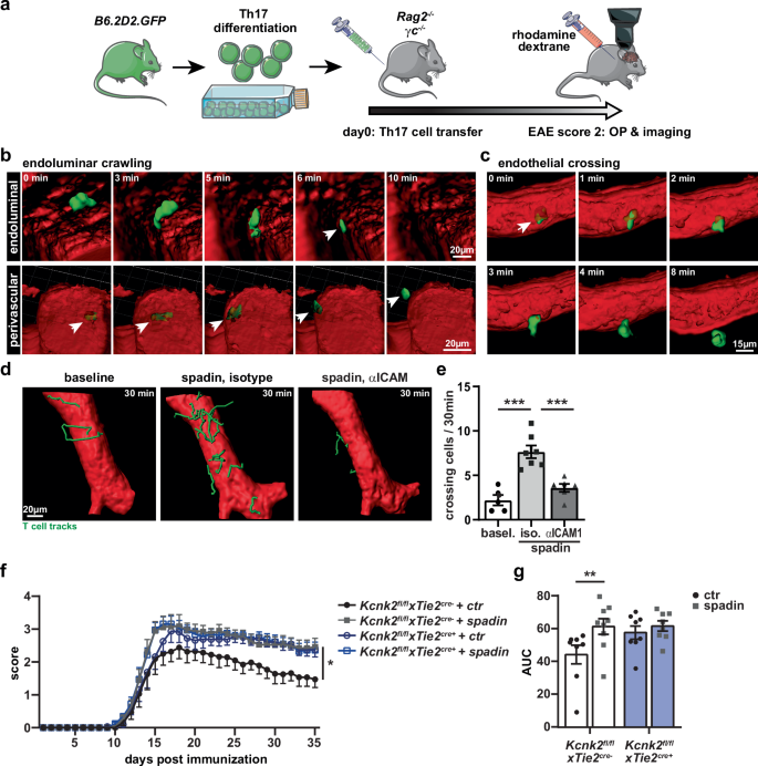
K2P2.1 (gene: Kcnk2), a two-pore-domain potassium channel, regulates leukocyte transmigration across the blood-brain barrier by a yet unknown mechanism. We demonstrate that Kcnk2−/− mouse brain microvascular endothelial cells (MBMECs) exhibit an altered cytoskeletal structure and surface morphology with increased formation of membrane protrusions. Cell adhesion molecules cluster on those protrusions and facilitate leukocyte adhesion and migration in vitro and in vivo. We observe downregulation of K2P2.1 and activation of actin modulating proteins (cofilin 1, Arp2/3) in inflamed wildtype MBMECs. In the mechanosensitive conformation, K2P2.1 shields the phospholipid PI(4,5)P2 from interaction with other actin regulatory proteins, especially cofilin 1. Consequently, after stimulus-related K2P2.1 downregulation and dislocation from PI(4,5)P2, actin rearrangements are induced. Thus, K2P2.1-mediated regulatory processes are essential for actin dynamics, fast, reversible, and pharmacologically targetable.
期刊介绍:
Nature Communications, an open-access journal, publishes high-quality research spanning all areas of the natural sciences. Papers featured in the journal showcase significant advances relevant to specialists in each respective field. With a 2-year impact factor of 16.6 (2022) and a median time of 8 days from submission to the first editorial decision, Nature Communications is committed to rapid dissemination of research findings. As a multidisciplinary journal, it welcomes contributions from biological, health, physical, chemical, Earth, social, mathematical, applied, and engineering sciences, aiming to highlight important breakthroughs within each domain.

 求助内容:
求助内容: 应助结果提醒方式:


