I I Vlasova, M D Yurkanova, A A Zolotopup, T O Klyucherev, P S Timashev
{"title":"一氧化氮供体对人巨噬细胞铁下垂的调节作用。","authors":"I I Vlasova, M D Yurkanova, A A Zolotopup, T O Klyucherev, P S Timashev","doi":"10.17691/stm2025.17.3.04","DOIUrl":null,"url":null,"abstract":"<p><p>Ferroptosis is a programmed form of cell death in which iron-dependent lipid peroxidation is the main feature. Macrophages are the major cells of the immune system, they function in a pro-oxidative environment, so the study of their susceptibility to ferroptosis and the search for approaches to its regulation are important. <b>The aim of the study</b> was to investigate ferroptosis in macrophages differentiated from THP-1 myeloid leukemia cells and to compare the effect of NO donors with different half-lives on the degree of ferroptosis development.</p><p><strong>Materials and methods: </strong>RSL3 and ML-162, inhibitors of glutathione peroxidase 4 (GPX4), and erastin, an inhibitor of cystine/ glutamate transport, were used to induce ferroptosis in THP-1 macrophages. The progression of ferroptosis was monitored using three independent methods: reduction of Alamar blue by live cells, measurement of lactate dehydrogenase in the medium, and the LIVE/DEAD assay. Ferroptotic cell death was proven by using the specific inhibitor ferrostatin-1 and by detecting lipid oxidation in cells using the BODIPY 581/591 C11 fluorescent probe.</p><p><strong>Results: </strong>RSL3 and ML-162 dose-dependently induced ferroptosis in cells. THP-1 macrophage ferroptosis is a slow process and begins ~5 h after inducer addition. Erastin was a weak ferroptosis inducer; however, it enhanced ferroptosis induced by GPX4 inhibitors. We compared the ability of two NO donors with different half-lives to affect THP-1 macrophage ferroptosis: DEA NONOate (2 min) and DTPA NONOate (3 h). Donors were added either once after the inducer at a concentration of 100-120 μM or repeatedly until reaching the final concentration. DEA had no effect on THP-1 macrophage ferroptosis, whereas DPTA completely inhibited ferroptosis.</p><p><strong>Conclusion: </strong>DTPA, being an NO donor with a half-life of 3 h at 37°С, can be used to inhibit ferroptosis in THP-1 macrophages, which develops within 17-19 h. Therefore, there are mechanisms of prolongation of NO action in cells that should be studied to use NO donors for regulation of cellular ferroptosis.</p>","PeriodicalId":520289,"journal":{"name":"Sovremennye tekhnologii v meditsine","volume":"17 3","pages":"41-48"},"PeriodicalIF":0.0000,"publicationDate":"2025-01-01","publicationTypes":"Journal Article","fieldsOfStudy":null,"isOpenAccess":false,"openAccessPdf":"https://www.ncbi.nlm.nih.gov/pmc/articles/PMC12261289/pdf/","citationCount":"0","resultStr":"{\"title\":\"Regulation of Ferroptosis in Human Macrophage by Nitric Oxide Donors.\",\"authors\":\"I I Vlasova, M D Yurkanova, A A Zolotopup, T O Klyucherev, P S Timashev\",\"doi\":\"10.17691/stm2025.17.3.04\",\"DOIUrl\":null,\"url\":null,\"abstract\":\"<p><p>Ferroptosis is a programmed form of cell death in which iron-dependent lipid peroxidation is the main feature. Macrophages are the major cells of the immune system, they function in a pro-oxidative environment, so the study of their susceptibility to ferroptosis and the search for approaches to its regulation are important. <b>The aim of the study</b> was to investigate ferroptosis in macrophages differentiated from THP-1 myeloid leukemia cells and to compare the effect of NO donors with different half-lives on the degree of ferroptosis development.</p><p><strong>Materials and methods: </strong>RSL3 and ML-162, inhibitors of glutathione peroxidase 4 (GPX4), and erastin, an inhibitor of cystine/ glutamate transport, were used to induce ferroptosis in THP-1 macrophages. The progression of ferroptosis was monitored using three independent methods: reduction of Alamar blue by live cells, measurement of lactate dehydrogenase in the medium, and the LIVE/DEAD assay. Ferroptotic cell death was proven by using the specific inhibitor ferrostatin-1 and by detecting lipid oxidation in cells using the BODIPY 581/591 C11 fluorescent probe.</p><p><strong>Results: </strong>RSL3 and ML-162 dose-dependently induced ferroptosis in cells. THP-1 macrophage ferroptosis is a slow process and begins ~5 h after inducer addition. Erastin was a weak ferroptosis inducer; however, it enhanced ferroptosis induced by GPX4 inhibitors. We compared the ability of two NO donors with different half-lives to affect THP-1 macrophage ferroptosis: DEA NONOate (2 min) and DTPA NONOate (3 h). Donors were added either once after the inducer at a concentration of 100-120 μM or repeatedly until reaching the final concentration. DEA had no effect on THP-1 macrophage ferroptosis, whereas DPTA completely inhibited ferroptosis.</p><p><strong>Conclusion: </strong>DTPA, being an NO donor with a half-life of 3 h at 37°С, can be used to inhibit ferroptosis in THP-1 macrophages, which develops within 17-19 h. Therefore, there are mechanisms of prolongation of NO action in cells that should be studied to use NO donors for regulation of cellular ferroptosis.</p>\",\"PeriodicalId\":520289,\"journal\":{\"name\":\"Sovremennye tekhnologii v meditsine\",\"volume\":\"17 3\",\"pages\":\"41-48\"},\"PeriodicalIF\":0.0000,\"publicationDate\":\"2025-01-01\",\"publicationTypes\":\"Journal Article\",\"fieldsOfStudy\":null,\"isOpenAccess\":false,\"openAccessPdf\":\"https://www.ncbi.nlm.nih.gov/pmc/articles/PMC12261289/pdf/\",\"citationCount\":\"0\",\"resultStr\":null,\"platform\":\"Semanticscholar\",\"paperid\":null,\"PeriodicalName\":\"Sovremennye tekhnologii v meditsine\",\"FirstCategoryId\":\"1085\",\"ListUrlMain\":\"https://doi.org/10.17691/stm2025.17.3.04\",\"RegionNum\":0,\"RegionCategory\":null,\"ArticlePicture\":[],\"TitleCN\":null,\"AbstractTextCN\":null,\"PMCID\":null,\"EPubDate\":\"2025/6/30 0:00:00\",\"PubModel\":\"Epub\",\"JCR\":\"\",\"JCRName\":\"\",\"Score\":null,\"Total\":0}","platform":"Semanticscholar","paperid":null,"PeriodicalName":"Sovremennye tekhnologii v meditsine","FirstCategoryId":"1085","ListUrlMain":"https://doi.org/10.17691/stm2025.17.3.04","RegionNum":0,"RegionCategory":null,"ArticlePicture":[],"TitleCN":null,"AbstractTextCN":null,"PMCID":null,"EPubDate":"2025/6/30 0:00:00","PubModel":"Epub","JCR":"","JCRName":"","Score":null,"Total":0}
Regulation of Ferroptosis in Human Macrophage by Nitric Oxide Donors.
Ferroptosis is a programmed form of cell death in which iron-dependent lipid peroxidation is the main feature. Macrophages are the major cells of the immune system, they function in a pro-oxidative environment, so the study of their susceptibility to ferroptosis and the search for approaches to its regulation are important. The aim of the study was to investigate ferroptosis in macrophages differentiated from THP-1 myeloid leukemia cells and to compare the effect of NO donors with different half-lives on the degree of ferroptosis development.
Materials and methods: RSL3 and ML-162, inhibitors of glutathione peroxidase 4 (GPX4), and erastin, an inhibitor of cystine/ glutamate transport, were used to induce ferroptosis in THP-1 macrophages. The progression of ferroptosis was monitored using three independent methods: reduction of Alamar blue by live cells, measurement of lactate dehydrogenase in the medium, and the LIVE/DEAD assay. Ferroptotic cell death was proven by using the specific inhibitor ferrostatin-1 and by detecting lipid oxidation in cells using the BODIPY 581/591 C11 fluorescent probe.
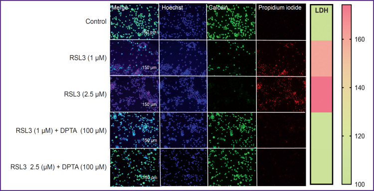
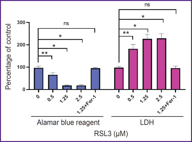
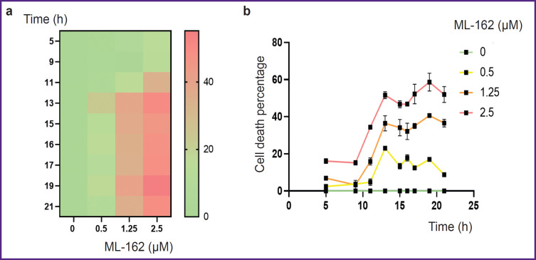
Results: RSL3 and ML-162 dose-dependently induced ferroptosis in cells. THP-1 macrophage ferroptosis is a slow process and begins ~5 h after inducer addition. Erastin was a weak ferroptosis inducer; however, it enhanced ferroptosis induced by GPX4 inhibitors. We compared the ability of two NO donors with different half-lives to affect THP-1 macrophage ferroptosis: DEA NONOate (2 min) and DTPA NONOate (3 h). Donors were added either once after the inducer at a concentration of 100-120 μM or repeatedly until reaching the final concentration. DEA had no effect on THP-1 macrophage ferroptosis, whereas DPTA completely inhibited ferroptosis.
Conclusion: DTPA, being an NO donor with a half-life of 3 h at 37°С, can be used to inhibit ferroptosis in THP-1 macrophages, which develops within 17-19 h. Therefore, there are mechanisms of prolongation of NO action in cells that should be studied to use NO donors for regulation of cellular ferroptosis.

 求助内容:
求助内容: 应助结果提醒方式:


