{"title":"急慢性闭角型青光眼晶状体位置及稳定性的超声生物显微成像分析。","authors":"Zhiying Yu, Xinyu Wang, Haitao Wang, Jing Han, Jing Fu, Licun Wang, Ling Wang","doi":"10.3389/fopht.2025.1624876","DOIUrl":null,"url":null,"abstract":"<p><strong>Introduction: </strong>This study aimed to compare the characteristics and differences in lens position and stability in patients with acute and chronic angle-closure glaucoma (ACG) using ultrasound biomicroscopy (UBM) to provide a basis for selecting treatment regimens for primary ACG (PACG).</p><p><strong>Methods: </strong>This prospective study included 82 eyes of patients with PACG, of which, 45 eyes with acute PACG (APACG), 37 with chronic PACG (CPACG). Axial length (AL) and lens thickness (LT) were measured using A-scan ultrasonography. Anterior chamber depth (ACD), pupil diameter (PD), and lens vault (LV) were measured using UBM for each group. Additionally, trabecular-iris angle (TIA), angle opening distance (AOD<sub>500</sub>), iris-lens angle (ILA), and iris-lens contact distance (ILCD) were measured in four quadrants (superior, inferior, nasal, and temporal) with UBM. The corresponding lens position (LP), relative lens position (RLP), and lens thickness/axial length factor (LAF) were calculated. Normally distributed data were compared between the two groups using an independent samples t-test. Data that did not follow a normal distribution were compared using the Mann-Whitney U test. Differences were considered statistically significant when <i>P</i> < 0.05, and they were considered highly statistically significant when <i>P</i> < 0.01.</p><p><strong>Results: </strong>The values for angle-related parameters, including the mean TIA, TIA<sub>max-min</sub>, mean AOD<sub>500</sub>, AOD<sub>500 max-min</sub>, and ACD, were significantly lower in the APACG group than in the CPACG group (all <i>P</i> < 0.05). The LP and RLP values of the APACG group were also lower than those of the CPACG group, but only the difference in LP values being statistically significant (<i>P</i> = 0.038). The LT, LV, LAF, mean ILCD, and ILCD<sub>max-min</sub> values were higher than those of the CPACG group, with the differences reaching statistical significance (all <i>P</i> < 0.05).</p><p><strong>Conclusion: </strong>The APACG eyes had a thicker and more-anteriorly positioned lens than those with CPACG, which results in a shallower anterior chamber and narrower anterior chamber angle. In the APACG group, the lens exhibited nonuniform laxity of the suspensory ligament across the various quadrants, poor stability, and greater susceptibility for anterior displacement or even deviation.</p>","PeriodicalId":73096,"journal":{"name":"Frontiers in ophthalmology","volume":"5 ","pages":"1624876"},"PeriodicalIF":0.9000,"publicationDate":"2025-07-02","publicationTypes":"Journal Article","fieldsOfStudy":null,"isOpenAccess":false,"openAccessPdf":"https://www.ncbi.nlm.nih.gov/pmc/articles/PMC12263399/pdf/","citationCount":"0","resultStr":"{\"title\":\"Ultrasound biomicroscopic imaging analysis of lens position and stability in acute and chronic angle-closure glaucoma.\",\"authors\":\"Zhiying Yu, Xinyu Wang, Haitao Wang, Jing Han, Jing Fu, Licun Wang, Ling Wang\",\"doi\":\"10.3389/fopht.2025.1624876\",\"DOIUrl\":null,\"url\":null,\"abstract\":\"<p><strong>Introduction: </strong>This study aimed to compare the characteristics and differences in lens position and stability in patients with acute and chronic angle-closure glaucoma (ACG) using ultrasound biomicroscopy (UBM) to provide a basis for selecting treatment regimens for primary ACG (PACG).</p><p><strong>Methods: </strong>This prospective study included 82 eyes of patients with PACG, of which, 45 eyes with acute PACG (APACG), 37 with chronic PACG (CPACG). Axial length (AL) and lens thickness (LT) were measured using A-scan ultrasonography. Anterior chamber depth (ACD), pupil diameter (PD), and lens vault (LV) were measured using UBM for each group. Additionally, trabecular-iris angle (TIA), angle opening distance (AOD<sub>500</sub>), iris-lens angle (ILA), and iris-lens contact distance (ILCD) were measured in four quadrants (superior, inferior, nasal, and temporal) with UBM. The corresponding lens position (LP), relative lens position (RLP), and lens thickness/axial length factor (LAF) were calculated. Normally distributed data were compared between the two groups using an independent samples t-test. Data that did not follow a normal distribution were compared using the Mann-Whitney U test. Differences were considered statistically significant when <i>P</i> < 0.05, and they were considered highly statistically significant when <i>P</i> < 0.01.</p><p><strong>Results: </strong>The values for angle-related parameters, including the mean TIA, TIA<sub>max-min</sub>, mean AOD<sub>500</sub>, AOD<sub>500 max-min</sub>, and ACD, were significantly lower in the APACG group than in the CPACG group (all <i>P</i> < 0.05). The LP and RLP values of the APACG group were also lower than those of the CPACG group, but only the difference in LP values being statistically significant (<i>P</i> = 0.038). The LT, LV, LAF, mean ILCD, and ILCD<sub>max-min</sub> values were higher than those of the CPACG group, with the differences reaching statistical significance (all <i>P</i> < 0.05).</p><p><strong>Conclusion: </strong>The APACG eyes had a thicker and more-anteriorly positioned lens than those with CPACG, which results in a shallower anterior chamber and narrower anterior chamber angle. In the APACG group, the lens exhibited nonuniform laxity of the suspensory ligament across the various quadrants, poor stability, and greater susceptibility for anterior displacement or even deviation.</p>\",\"PeriodicalId\":73096,\"journal\":{\"name\":\"Frontiers in ophthalmology\",\"volume\":\"5 \",\"pages\":\"1624876\"},\"PeriodicalIF\":0.9000,\"publicationDate\":\"2025-07-02\",\"publicationTypes\":\"Journal Article\",\"fieldsOfStudy\":null,\"isOpenAccess\":false,\"openAccessPdf\":\"https://www.ncbi.nlm.nih.gov/pmc/articles/PMC12263399/pdf/\",\"citationCount\":\"0\",\"resultStr\":null,\"platform\":\"Semanticscholar\",\"paperid\":null,\"PeriodicalName\":\"Frontiers in ophthalmology\",\"FirstCategoryId\":\"1085\",\"ListUrlMain\":\"https://doi.org/10.3389/fopht.2025.1624876\",\"RegionNum\":0,\"RegionCategory\":null,\"ArticlePicture\":[],\"TitleCN\":null,\"AbstractTextCN\":null,\"PMCID\":null,\"EPubDate\":\"2025/1/1 0:00:00\",\"PubModel\":\"eCollection\",\"JCR\":\"\",\"JCRName\":\"\",\"Score\":null,\"Total\":0}","platform":"Semanticscholar","paperid":null,"PeriodicalName":"Frontiers in ophthalmology","FirstCategoryId":"1085","ListUrlMain":"https://doi.org/10.3389/fopht.2025.1624876","RegionNum":0,"RegionCategory":null,"ArticlePicture":[],"TitleCN":null,"AbstractTextCN":null,"PMCID":null,"EPubDate":"2025/1/1 0:00:00","PubModel":"eCollection","JCR":"","JCRName":"","Score":null,"Total":0}
Ultrasound biomicroscopic imaging analysis of lens position and stability in acute and chronic angle-closure glaucoma.
Introduction: This study aimed to compare the characteristics and differences in lens position and stability in patients with acute and chronic angle-closure glaucoma (ACG) using ultrasound biomicroscopy (UBM) to provide a basis for selecting treatment regimens for primary ACG (PACG).
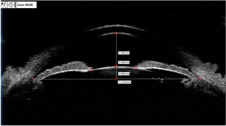
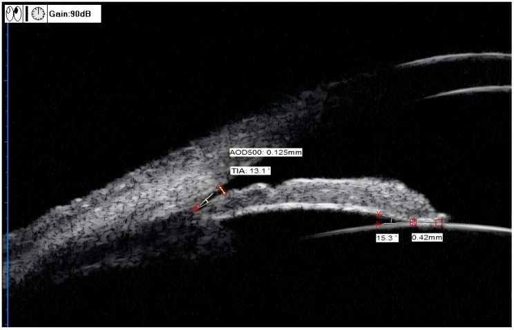
Methods: This prospective study included 82 eyes of patients with PACG, of which, 45 eyes with acute PACG (APACG), 37 with chronic PACG (CPACG). Axial length (AL) and lens thickness (LT) were measured using A-scan ultrasonography. Anterior chamber depth (ACD), pupil diameter (PD), and lens vault (LV) were measured using UBM for each group. Additionally, trabecular-iris angle (TIA), angle opening distance (AOD500), iris-lens angle (ILA), and iris-lens contact distance (ILCD) were measured in four quadrants (superior, inferior, nasal, and temporal) with UBM. The corresponding lens position (LP), relative lens position (RLP), and lens thickness/axial length factor (LAF) were calculated. Normally distributed data were compared between the two groups using an independent samples t-test. Data that did not follow a normal distribution were compared using the Mann-Whitney U test. Differences were considered statistically significant when P < 0.05, and they were considered highly statistically significant when P < 0.01.
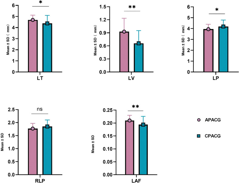
Results: The values for angle-related parameters, including the mean TIA, TIAmax-min, mean AOD500, AOD500 max-min, and ACD, were significantly lower in the APACG group than in the CPACG group (all P < 0.05). The LP and RLP values of the APACG group were also lower than those of the CPACG group, but only the difference in LP values being statistically significant (P = 0.038). The LT, LV, LAF, mean ILCD, and ILCDmax-min values were higher than those of the CPACG group, with the differences reaching statistical significance (all P < 0.05).
Conclusion: The APACG eyes had a thicker and more-anteriorly positioned lens than those with CPACG, which results in a shallower anterior chamber and narrower anterior chamber angle. In the APACG group, the lens exhibited nonuniform laxity of the suspensory ligament across the various quadrants, poor stability, and greater susceptibility for anterior displacement or even deviation.

 求助内容:
求助内容: 应助结果提醒方式:


