Hung-Da Chou, Rodrigo Anguita, Caroline Thaung, Lamis Alharby, Guy S Negretti, Lyndon da Cruz, Mandeep S Sagoo
{"title":"23号玻璃体诊断活检后葡萄膜黑色素瘤的播散。","authors":"Hung-Da Chou, Rodrigo Anguita, Caroline Thaung, Lamis Alharby, Guy S Negretti, Lyndon da Cruz, Mandeep S Sagoo","doi":"10.1097/IAE.0000000000004573","DOIUrl":null,"url":null,"abstract":"<p><strong>Purpose: </strong>To report a case with tumor dissemination after diagnostic choroidal biopsy.</p><p><strong>Methods: </strong>Multimodal imaging and histopathology correlation.</p><p><strong>Results: </strong>A 64-year-old male presented with an atypical uveal mass and underwent a diagnostic transretinal 23-gauge vitrector biopsy to diagnose melanoma. Post-operative course was complicated by prolonged vitreous hemorrhage which required vitrectomy, delaying proton beam treatment by two months. One year after the biopsy, the tumor cells had disseminated to the retina, optic nerve, iris, iridocorneal angle, and extraocularly via the vitrectomy port. The patient died from metastatic melanoma one year later. To the best of our knowledge, there are 10 prior cases with biopsy-related tumor seeding in the literature, with risk features including multiple vitreous surgeries, a long interval between biopsy and treatment, and high-risk melanoma cytogenetics. Severe vitreous hemorrhage following biopsy further postpone treatments or warrant vitrectomy, both increasing the risk of tumor spread.</p><p><strong>Conclusions: </strong>Biopsy of choroidal melanoma, especially when complicated by severe vitreous hemorrhage, can lead to dissemination of the malignant tumor.</p>","PeriodicalId":54486,"journal":{"name":"Retina-The Journal of Retinal and Vitreous Diseases","volume":" ","pages":""},"PeriodicalIF":2.1000,"publicationDate":"2025-07-07","publicationTypes":"Journal Article","fieldsOfStudy":null,"isOpenAccess":false,"openAccessPdf":"https://www.ncbi.nlm.nih.gov/pmc/articles/PMC12366731/pdf/","citationCount":"0","resultStr":"{\"title\":\"Dissemination of Uveal Melanoma After Diagnostic Biopsy With 23-Gauge Vitrector.\",\"authors\":\"Hung-Da Chou, Rodrigo Anguita, Caroline Thaung, Lamis Alharby, Guy S Negretti, Lyndon da Cruz, Mandeep S Sagoo\",\"doi\":\"10.1097/IAE.0000000000004573\",\"DOIUrl\":null,\"url\":null,\"abstract\":\"<p><strong>Purpose: </strong>To report a case with tumor dissemination after diagnostic choroidal biopsy.</p><p><strong>Methods: </strong>Multimodal imaging and histopathology correlation.</p><p><strong>Results: </strong>A 64-year-old male presented with an atypical uveal mass and underwent a diagnostic transretinal 23-gauge vitrector biopsy to diagnose melanoma. Post-operative course was complicated by prolonged vitreous hemorrhage which required vitrectomy, delaying proton beam treatment by two months. One year after the biopsy, the tumor cells had disseminated to the retina, optic nerve, iris, iridocorneal angle, and extraocularly via the vitrectomy port. The patient died from metastatic melanoma one year later. To the best of our knowledge, there are 10 prior cases with biopsy-related tumor seeding in the literature, with risk features including multiple vitreous surgeries, a long interval between biopsy and treatment, and high-risk melanoma cytogenetics. Severe vitreous hemorrhage following biopsy further postpone treatments or warrant vitrectomy, both increasing the risk of tumor spread.</p><p><strong>Conclusions: </strong>Biopsy of choroidal melanoma, especially when complicated by severe vitreous hemorrhage, can lead to dissemination of the malignant tumor.</p>\",\"PeriodicalId\":54486,\"journal\":{\"name\":\"Retina-The Journal of Retinal and Vitreous Diseases\",\"volume\":\" \",\"pages\":\"\"},\"PeriodicalIF\":2.1000,\"publicationDate\":\"2025-07-07\",\"publicationTypes\":\"Journal Article\",\"fieldsOfStudy\":null,\"isOpenAccess\":false,\"openAccessPdf\":\"https://www.ncbi.nlm.nih.gov/pmc/articles/PMC12366731/pdf/\",\"citationCount\":\"0\",\"resultStr\":null,\"platform\":\"Semanticscholar\",\"paperid\":null,\"PeriodicalName\":\"Retina-The Journal of Retinal and Vitreous Diseases\",\"FirstCategoryId\":\"3\",\"ListUrlMain\":\"https://doi.org/10.1097/IAE.0000000000004573\",\"RegionNum\":2,\"RegionCategory\":\"医学\",\"ArticlePicture\":[],\"TitleCN\":null,\"AbstractTextCN\":null,\"PMCID\":null,\"EPubDate\":\"\",\"PubModel\":\"\",\"JCR\":\"Q2\",\"JCRName\":\"OPHTHALMOLOGY\",\"Score\":null,\"Total\":0}","platform":"Semanticscholar","paperid":null,"PeriodicalName":"Retina-The Journal of Retinal and Vitreous Diseases","FirstCategoryId":"3","ListUrlMain":"https://doi.org/10.1097/IAE.0000000000004573","RegionNum":2,"RegionCategory":"医学","ArticlePicture":[],"TitleCN":null,"AbstractTextCN":null,"PMCID":null,"EPubDate":"","PubModel":"","JCR":"Q2","JCRName":"OPHTHALMOLOGY","Score":null,"Total":0}
Dissemination of Uveal Melanoma After Diagnostic Biopsy With 23-Gauge Vitrector.
Purpose: To report a case with tumor dissemination after diagnostic choroidal biopsy.
Methods: Multimodal imaging and histopathology correlation.
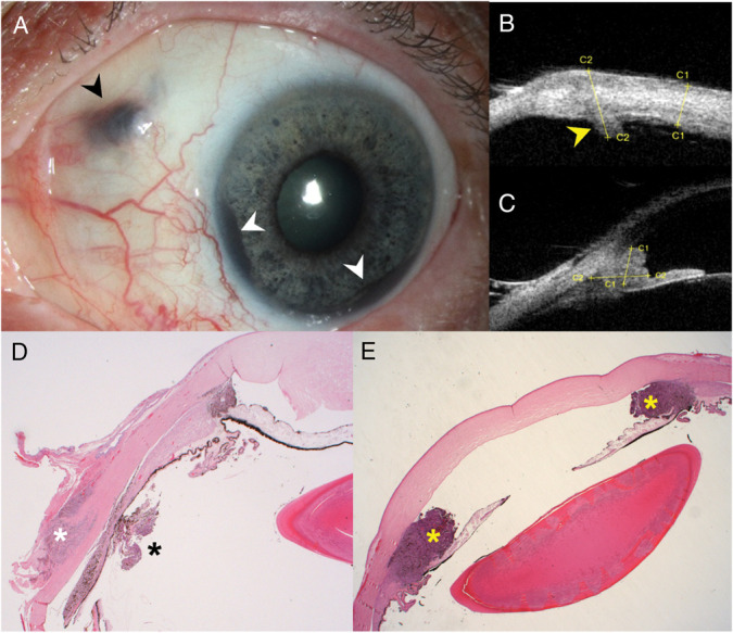
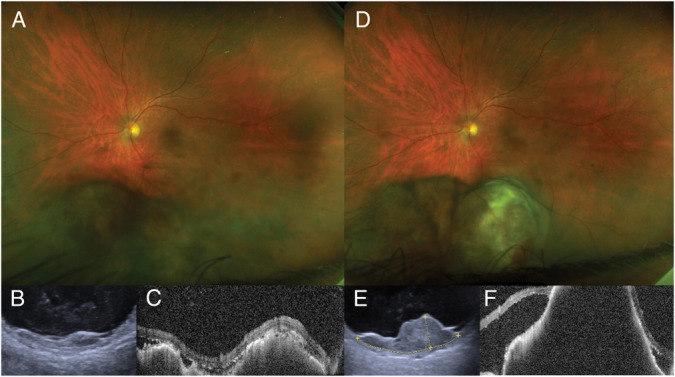
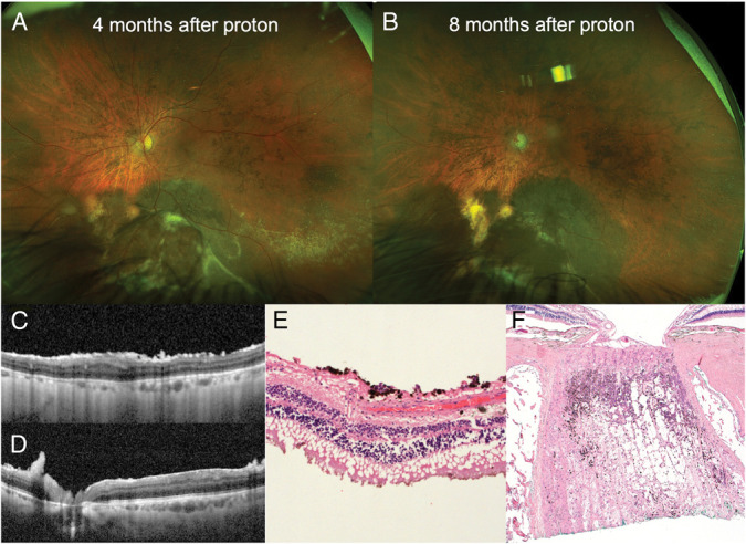
Results: A 64-year-old male presented with an atypical uveal mass and underwent a diagnostic transretinal 23-gauge vitrector biopsy to diagnose melanoma. Post-operative course was complicated by prolonged vitreous hemorrhage which required vitrectomy, delaying proton beam treatment by two months. One year after the biopsy, the tumor cells had disseminated to the retina, optic nerve, iris, iridocorneal angle, and extraocularly via the vitrectomy port. The patient died from metastatic melanoma one year later. To the best of our knowledge, there are 10 prior cases with biopsy-related tumor seeding in the literature, with risk features including multiple vitreous surgeries, a long interval between biopsy and treatment, and high-risk melanoma cytogenetics. Severe vitreous hemorrhage following biopsy further postpone treatments or warrant vitrectomy, both increasing the risk of tumor spread.
Conclusions: Biopsy of choroidal melanoma, especially when complicated by severe vitreous hemorrhage, can lead to dissemination of the malignant tumor.
期刊介绍:
RETINA® focuses exclusively on the growing specialty of vitreoretinal disorders. The Journal provides current information on diagnostic and therapeutic techniques. Its highly specialized and informative, peer-reviewed articles are easily applicable to clinical practice.
In addition to regular reports from clinical and basic science investigators, RETINA® publishes special features including periodic review articles on pertinent topics, special articles dealing with surgical and other therapeutic techniques, and abstract cards. Issues are abundantly illustrated in vivid full color.
Published 12 times per year, RETINA® is truly a “must have” publication for anyone connected to this field.

 求助内容:
求助内容: 应助结果提醒方式:


