{"title":"通过放射组学增强术中对浸润性肺腺癌高级别模式的识别。","authors":"Yuanxin Sun, Hao Dong, Weiqiu Jin, Haoxiang Xuan, Zheng Yuan, Lukas Käsmann, Leilei Shen, Tingting Wang, Xiaodan Ye, Mengsu Zeng","doi":"10.21037/tlcr-2025-504","DOIUrl":null,"url":null,"abstract":"<p><strong>Background: </strong>High-grade patterns (HGPs) are important for surgical decision-making in patients with invasive lung adenocarcinoma (IAC), but the sensitivity of intraoperative frozen section (FS) is not high. Radiomics has the potential to improve the sensitivity of intraoperative detection. The purpose of the present study was to evaluate the value of combining radiomics with FS analysis for predicting HGPs in patients with clinical T1 (cT1) IAC.</p><p><strong>Methods: </strong>Data from a total of 490 patients who were surgically diagnosed with IAC from January 2019 to April 2019 were retrospectively analyzed; the patients were randomly divided into a training set (n=392) and a test set (n=98). The presence of HGPs (micropapillary, solid, and complex glandular patterns) was evaluated according to the final pathology (FP). Radiomics features were extracted from thin-slice computed tomography (CT) images, and feature selection was performed via the mutual information method and least absolute shrinkage and selection operator regression algorithm. The radiomics (R), FS, and radiomics-frozen section (R-FS) models were established to predict the presence of HGPs in FP. The area under the receiver operating characteristic (ROC) curve, the precision-recall curve, the calibration curve, and decision curve analysis were used to evaluate model performances. The permutation importance algorithm (PIA) and local interpretable model-agnostic explanations (LIME) were used to provide interpretations for the R model. Additionally, the predictive performance was compared among tumors with different CT densities.</p><p><strong>Results: </strong>The R and R-FS models outperformed the FS model, with the R-FS model achieving the best area under the curve value of 0.907 (95% confidence interval: 0.830-0.956) in the test set. PIA and LIME determined the interpretability of outputs from both the overall model and individual sample perspectives. Among the three models, the R model performed best in pure ground-glass nodules and pure-solid tumors.</p><p><strong>Conclusions: </strong>Radiomics could function as a complementary check to FS to provide a more sensible and accurate intraoperative identification of HGPs as compared to the use of FS alone, thus better informing clinical decision-making.</p>","PeriodicalId":23271,"journal":{"name":"Translational lung cancer research","volume":"14 6","pages":"2145-2158"},"PeriodicalIF":3.5000,"publicationDate":"2025-06-30","publicationTypes":"Journal Article","fieldsOfStudy":null,"isOpenAccess":false,"openAccessPdf":"https://www.ncbi.nlm.nih.gov/pmc/articles/PMC12261232/pdf/","citationCount":"0","resultStr":"{\"title\":\"Enhancing the intraoperative identification of high-grade patterns in invasive lung adenocarcinoma via radiomics.\",\"authors\":\"Yuanxin Sun, Hao Dong, Weiqiu Jin, Haoxiang Xuan, Zheng Yuan, Lukas Käsmann, Leilei Shen, Tingting Wang, Xiaodan Ye, Mengsu Zeng\",\"doi\":\"10.21037/tlcr-2025-504\",\"DOIUrl\":null,\"url\":null,\"abstract\":\"<p><strong>Background: </strong>High-grade patterns (HGPs) are important for surgical decision-making in patients with invasive lung adenocarcinoma (IAC), but the sensitivity of intraoperative frozen section (FS) is not high. Radiomics has the potential to improve the sensitivity of intraoperative detection. The purpose of the present study was to evaluate the value of combining radiomics with FS analysis for predicting HGPs in patients with clinical T1 (cT1) IAC.</p><p><strong>Methods: </strong>Data from a total of 490 patients who were surgically diagnosed with IAC from January 2019 to April 2019 were retrospectively analyzed; the patients were randomly divided into a training set (n=392) and a test set (n=98). The presence of HGPs (micropapillary, solid, and complex glandular patterns) was evaluated according to the final pathology (FP). Radiomics features were extracted from thin-slice computed tomography (CT) images, and feature selection was performed via the mutual information method and least absolute shrinkage and selection operator regression algorithm. The radiomics (R), FS, and radiomics-frozen section (R-FS) models were established to predict the presence of HGPs in FP. The area under the receiver operating characteristic (ROC) curve, the precision-recall curve, the calibration curve, and decision curve analysis were used to evaluate model performances. The permutation importance algorithm (PIA) and local interpretable model-agnostic explanations (LIME) were used to provide interpretations for the R model. Additionally, the predictive performance was compared among tumors with different CT densities.</p><p><strong>Results: </strong>The R and R-FS models outperformed the FS model, with the R-FS model achieving the best area under the curve value of 0.907 (95% confidence interval: 0.830-0.956) in the test set. PIA and LIME determined the interpretability of outputs from both the overall model and individual sample perspectives. Among the three models, the R model performed best in pure ground-glass nodules and pure-solid tumors.</p><p><strong>Conclusions: </strong>Radiomics could function as a complementary check to FS to provide a more sensible and accurate intraoperative identification of HGPs as compared to the use of FS alone, thus better informing clinical decision-making.</p>\",\"PeriodicalId\":23271,\"journal\":{\"name\":\"Translational lung cancer research\",\"volume\":\"14 6\",\"pages\":\"2145-2158\"},\"PeriodicalIF\":3.5000,\"publicationDate\":\"2025-06-30\",\"publicationTypes\":\"Journal Article\",\"fieldsOfStudy\":null,\"isOpenAccess\":false,\"openAccessPdf\":\"https://www.ncbi.nlm.nih.gov/pmc/articles/PMC12261232/pdf/\",\"citationCount\":\"0\",\"resultStr\":null,\"platform\":\"Semanticscholar\",\"paperid\":null,\"PeriodicalName\":\"Translational lung cancer research\",\"FirstCategoryId\":\"3\",\"ListUrlMain\":\"https://doi.org/10.21037/tlcr-2025-504\",\"RegionNum\":2,\"RegionCategory\":\"医学\",\"ArticlePicture\":[],\"TitleCN\":null,\"AbstractTextCN\":null,\"PMCID\":null,\"EPubDate\":\"2025/6/26 0:00:00\",\"PubModel\":\"Epub\",\"JCR\":\"Q2\",\"JCRName\":\"ONCOLOGY\",\"Score\":null,\"Total\":0}","platform":"Semanticscholar","paperid":null,"PeriodicalName":"Translational lung cancer research","FirstCategoryId":"3","ListUrlMain":"https://doi.org/10.21037/tlcr-2025-504","RegionNum":2,"RegionCategory":"医学","ArticlePicture":[],"TitleCN":null,"AbstractTextCN":null,"PMCID":null,"EPubDate":"2025/6/26 0:00:00","PubModel":"Epub","JCR":"Q2","JCRName":"ONCOLOGY","Score":null,"Total":0}
Enhancing the intraoperative identification of high-grade patterns in invasive lung adenocarcinoma via radiomics.
Background: High-grade patterns (HGPs) are important for surgical decision-making in patients with invasive lung adenocarcinoma (IAC), but the sensitivity of intraoperative frozen section (FS) is not high. Radiomics has the potential to improve the sensitivity of intraoperative detection. The purpose of the present study was to evaluate the value of combining radiomics with FS analysis for predicting HGPs in patients with clinical T1 (cT1) IAC.
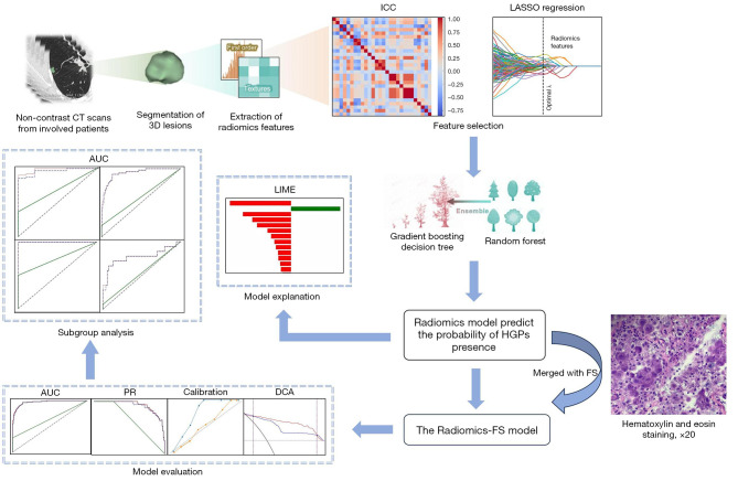
Methods: Data from a total of 490 patients who were surgically diagnosed with IAC from January 2019 to April 2019 were retrospectively analyzed; the patients were randomly divided into a training set (n=392) and a test set (n=98). The presence of HGPs (micropapillary, solid, and complex glandular patterns) was evaluated according to the final pathology (FP). Radiomics features were extracted from thin-slice computed tomography (CT) images, and feature selection was performed via the mutual information method and least absolute shrinkage and selection operator regression algorithm. The radiomics (R), FS, and radiomics-frozen section (R-FS) models were established to predict the presence of HGPs in FP. The area under the receiver operating characteristic (ROC) curve, the precision-recall curve, the calibration curve, and decision curve analysis were used to evaluate model performances. The permutation importance algorithm (PIA) and local interpretable model-agnostic explanations (LIME) were used to provide interpretations for the R model. Additionally, the predictive performance was compared among tumors with different CT densities.
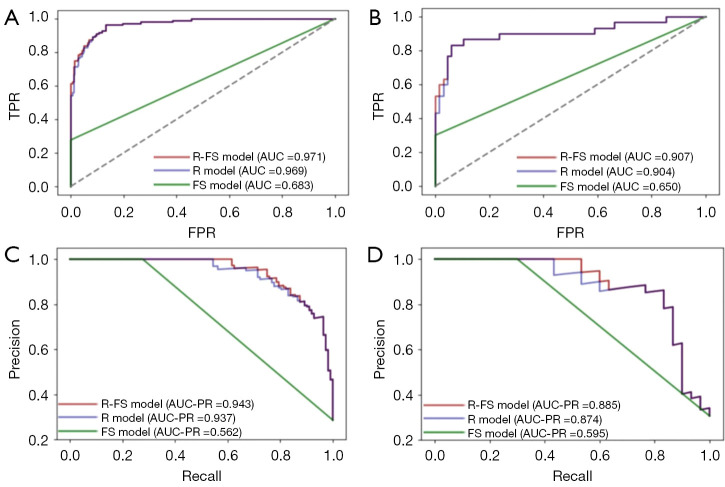
Results: The R and R-FS models outperformed the FS model, with the R-FS model achieving the best area under the curve value of 0.907 (95% confidence interval: 0.830-0.956) in the test set. PIA and LIME determined the interpretability of outputs from both the overall model and individual sample perspectives. Among the three models, the R model performed best in pure ground-glass nodules and pure-solid tumors.
Conclusions: Radiomics could function as a complementary check to FS to provide a more sensible and accurate intraoperative identification of HGPs as compared to the use of FS alone, thus better informing clinical decision-making.
期刊介绍:
Translational Lung Cancer Research(TLCR, Transl Lung Cancer Res, Print ISSN 2218-6751; Online ISSN 2226-4477) is an international, peer-reviewed, open-access journal, which was founded in March 2012. TLCR is indexed by PubMed/PubMed Central and the Chemical Abstracts Service (CAS) Databases. It is published quarterly the first year, and published bimonthly since February 2013. It provides practical up-to-date information on prevention, early detection, diagnosis, and treatment of lung cancer. Specific areas of its interest include, but not limited to, multimodality therapy, markers, imaging, tumor biology, pathology, chemoprevention, and technical advances related to lung cancer.

 求助内容:
求助内容: 应助结果提醒方式:


