Florian Raab, Quirin Strotzer, Christian Stroszczynski, Claudia Fellner, Ingo Einspieler, Michael Haimerl, Elmar W Lang
{"title":"基于nnU-Net和Swin UNETR的多期MRI肝脏结构自动分割。","authors":"Florian Raab, Quirin Strotzer, Christian Stroszczynski, Claudia Fellner, Ingo Einspieler, Michael Haimerl, Elmar W Lang","doi":"10.1038/s41598-025-07084-5","DOIUrl":null,"url":null,"abstract":"<p><p>Accurate segmentation of the liver parenchyma, portal veins, hepatic veins, and lesions from MRI is important for hepatic disease monitoring and treatment. Multi-phase contrast enhanced imaging is superior in distinguishing hepatic structures compared to single-phase approaches, but automated approaches for detailed segmentation of hepatic structures are lacking. This study evaluates deep learning architectures for segmenting liver structures from multi-phase Gd-EOB-DTPA-enhanced T1-weighted VIBE MRI scans. We utilized 458 T1-weighted VIBE scans of pathological livers, with 78 manually labeled for liver parenchyma, hepatic and portal veins, aorta, lesions, and ascites. An additional dataset of 47 labeled subjects was used for cross-scanner evaluation. Three models were evaluated using nested cross-validation: the conventional nnU-Net, the ResEnc nnU-Net, and the Swin UNETR. The late arterial phase was identified as the optimal fixed phase for co-registration. Both nnU-Net variants outperformed Swin UNETR across most tasks. The conventional nnU-Net achieved the highest segmentation performance for liver parenchyma (DSC: 0.97; 95% CI 0.97, 0.98), portal vein (DSC: 0.83; 95% CI 0.80, 0.87), and hepatic vein (DSC: 0.78; 95% CI 0.77, 0.80). Lesion and ascites segmentation proved challenging for all models, with the conventional nnU-Net performing best. This study demonstrates the effectiveness of deep learning, particularly nnU-Net variants, for detailed liver structure segmentation from multi-phase MRI. The developed models and preprocessing pipeline offer potential for improved liver disease assessment and surgical planning in clinical practice.</p>","PeriodicalId":21811,"journal":{"name":"Scientific Reports","volume":"15 1","pages":"25740"},"PeriodicalIF":3.9000,"publicationDate":"2025-07-16","publicationTypes":"Journal Article","fieldsOfStudy":null,"isOpenAccess":false,"openAccessPdf":"https://www.ncbi.nlm.nih.gov/pmc/articles/PMC12267559/pdf/","citationCount":"0","resultStr":"{\"title\":\"Automatic segmentation of liver structures in multi-phase MRI using variants of nnU-Net and Swin UNETR.\",\"authors\":\"Florian Raab, Quirin Strotzer, Christian Stroszczynski, Claudia Fellner, Ingo Einspieler, Michael Haimerl, Elmar W Lang\",\"doi\":\"10.1038/s41598-025-07084-5\",\"DOIUrl\":null,\"url\":null,\"abstract\":\"<p><p>Accurate segmentation of the liver parenchyma, portal veins, hepatic veins, and lesions from MRI is important for hepatic disease monitoring and treatment. Multi-phase contrast enhanced imaging is superior in distinguishing hepatic structures compared to single-phase approaches, but automated approaches for detailed segmentation of hepatic structures are lacking. This study evaluates deep learning architectures for segmenting liver structures from multi-phase Gd-EOB-DTPA-enhanced T1-weighted VIBE MRI scans. We utilized 458 T1-weighted VIBE scans of pathological livers, with 78 manually labeled for liver parenchyma, hepatic and portal veins, aorta, lesions, and ascites. An additional dataset of 47 labeled subjects was used for cross-scanner evaluation. Three models were evaluated using nested cross-validation: the conventional nnU-Net, the ResEnc nnU-Net, and the Swin UNETR. The late arterial phase was identified as the optimal fixed phase for co-registration. Both nnU-Net variants outperformed Swin UNETR across most tasks. The conventional nnU-Net achieved the highest segmentation performance for liver parenchyma (DSC: 0.97; 95% CI 0.97, 0.98), portal vein (DSC: 0.83; 95% CI 0.80, 0.87), and hepatic vein (DSC: 0.78; 95% CI 0.77, 0.80). Lesion and ascites segmentation proved challenging for all models, with the conventional nnU-Net performing best. This study demonstrates the effectiveness of deep learning, particularly nnU-Net variants, for detailed liver structure segmentation from multi-phase MRI. The developed models and preprocessing pipeline offer potential for improved liver disease assessment and surgical planning in clinical practice.</p>\",\"PeriodicalId\":21811,\"journal\":{\"name\":\"Scientific Reports\",\"volume\":\"15 1\",\"pages\":\"25740\"},\"PeriodicalIF\":3.9000,\"publicationDate\":\"2025-07-16\",\"publicationTypes\":\"Journal Article\",\"fieldsOfStudy\":null,\"isOpenAccess\":false,\"openAccessPdf\":\"https://www.ncbi.nlm.nih.gov/pmc/articles/PMC12267559/pdf/\",\"citationCount\":\"0\",\"resultStr\":null,\"platform\":\"Semanticscholar\",\"paperid\":null,\"PeriodicalName\":\"Scientific Reports\",\"FirstCategoryId\":\"103\",\"ListUrlMain\":\"https://doi.org/10.1038/s41598-025-07084-5\",\"RegionNum\":2,\"RegionCategory\":\"综合性期刊\",\"ArticlePicture\":[],\"TitleCN\":null,\"AbstractTextCN\":null,\"PMCID\":null,\"EPubDate\":\"\",\"PubModel\":\"\",\"JCR\":\"Q1\",\"JCRName\":\"MULTIDISCIPLINARY SCIENCES\",\"Score\":null,\"Total\":0}","platform":"Semanticscholar","paperid":null,"PeriodicalName":"Scientific Reports","FirstCategoryId":"103","ListUrlMain":"https://doi.org/10.1038/s41598-025-07084-5","RegionNum":2,"RegionCategory":"综合性期刊","ArticlePicture":[],"TitleCN":null,"AbstractTextCN":null,"PMCID":null,"EPubDate":"","PubModel":"","JCR":"Q1","JCRName":"MULTIDISCIPLINARY SCIENCES","Score":null,"Total":0}
引用次数: 0
摘要
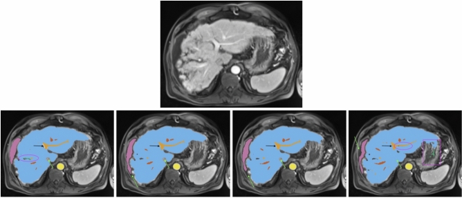
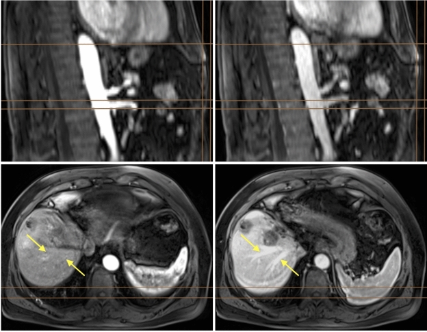
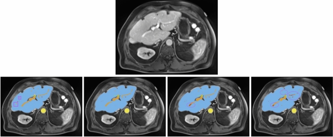
MRI准确分割肝实质、门静脉、肝静脉和病变对肝病监测和治疗非常重要。与单相方法相比,多相对比增强成像在区分肝脏结构方面优于单相方法,但缺乏详细分割肝脏结构的自动化方法。本研究评估了深度学习架构在多相gd - eob - dtpa增强t1加权VIBE MRI扫描中分割肝脏结构的效果。我们使用了458张病理肝脏的t1加权VIBE扫描,其中78张手工标记了肝实质、肝静脉和门静脉、主动脉、病变和腹水。另外47个被标记对象的数据集用于交叉扫描仪评估。使用嵌套交叉验证对三个模型进行评估:传统的nnU-Net, ResEnc的nnU-Net和Swin UNETR。确定动脉晚期为共登记的最佳固定期。两种nnU-Net变体在大多数任务上都优于Swin UNETR。传统的nnU-Net对肝实质的分割效果最好(DSC: 0.97;95% CI 0.97, 0.98),门静脉(DSC: 0.83;95% CI 0.80, 0.87)和肝静脉(DSC: 0.78;95% ci 0.77, 0.80)。病变和腹水分割对所有模型都具有挑战性,传统的nnU-Net表现最好。这项研究证明了深度学习的有效性,特别是nnU-Net变体,可以从多期MRI中进行详细的肝脏结构分割。开发的模型和预处理管道为临床实践中改善肝脏疾病评估和手术计划提供了潜力。
Automatic segmentation of liver structures in multi-phase MRI using variants of nnU-Net and Swin UNETR.
Accurate segmentation of the liver parenchyma, portal veins, hepatic veins, and lesions from MRI is important for hepatic disease monitoring and treatment. Multi-phase contrast enhanced imaging is superior in distinguishing hepatic structures compared to single-phase approaches, but automated approaches for detailed segmentation of hepatic structures are lacking. This study evaluates deep learning architectures for segmenting liver structures from multi-phase Gd-EOB-DTPA-enhanced T1-weighted VIBE MRI scans. We utilized 458 T1-weighted VIBE scans of pathological livers, with 78 manually labeled for liver parenchyma, hepatic and portal veins, aorta, lesions, and ascites. An additional dataset of 47 labeled subjects was used for cross-scanner evaluation. Three models were evaluated using nested cross-validation: the conventional nnU-Net, the ResEnc nnU-Net, and the Swin UNETR. The late arterial phase was identified as the optimal fixed phase for co-registration. Both nnU-Net variants outperformed Swin UNETR across most tasks. The conventional nnU-Net achieved the highest segmentation performance for liver parenchyma (DSC: 0.97; 95% CI 0.97, 0.98), portal vein (DSC: 0.83; 95% CI 0.80, 0.87), and hepatic vein (DSC: 0.78; 95% CI 0.77, 0.80). Lesion and ascites segmentation proved challenging for all models, with the conventional nnU-Net performing best. This study demonstrates the effectiveness of deep learning, particularly nnU-Net variants, for detailed liver structure segmentation from multi-phase MRI. The developed models and preprocessing pipeline offer potential for improved liver disease assessment and surgical planning in clinical practice.
期刊介绍:
We publish original research from all areas of the natural sciences, psychology, medicine and engineering. You can learn more about what we publish by browsing our specific scientific subject areas below or explore Scientific Reports by browsing all articles and collections.
Scientific Reports has a 2-year impact factor: 4.380 (2021), and is the 6th most-cited journal in the world, with more than 540,000 citations in 2020 (Clarivate Analytics, 2021).
•Engineering
Engineering covers all aspects of engineering, technology, and applied science. It plays a crucial role in the development of technologies to address some of the world''s biggest challenges, helping to save lives and improve the way we live.
•Physical sciences
Physical sciences are those academic disciplines that aim to uncover the underlying laws of nature — often written in the language of mathematics. It is a collective term for areas of study including astronomy, chemistry, materials science and physics.
•Earth and environmental sciences
Earth and environmental sciences cover all aspects of Earth and planetary science and broadly encompass solid Earth processes, surface and atmospheric dynamics, Earth system history, climate and climate change, marine and freshwater systems, and ecology. It also considers the interactions between humans and these systems.
•Biological sciences
Biological sciences encompass all the divisions of natural sciences examining various aspects of vital processes. The concept includes anatomy, physiology, cell biology, biochemistry and biophysics, and covers all organisms from microorganisms, animals to plants.
•Health sciences
The health sciences study health, disease and healthcare. This field of study aims to develop knowledge, interventions and technology for use in healthcare to improve the treatment of patients.
 求助内容:
求助内容: 应助结果提醒方式:


