Khawaja B Waheed, Ali Al Orf, Faisal Al Zahrani, Muhammad Z Ulhassan, Nawaf N Al Jubair, Zechariah J Arulanatham
{"title":"敏感性增强磁共振成像检测癫痫患者静脉血管瘤。","authors":"Khawaja B Waheed, Ali Al Orf, Faisal Al Zahrani, Muhammad Z Ulhassan, Nawaf N Al Jubair, Zechariah J Arulanatham","doi":"10.17712/nsj.2025.3.20240103","DOIUrl":null,"url":null,"abstract":"<p><strong>Objectives: </strong>To evaluate the use of susceptibility-enhanced sequence with conventional magnetic resonance imaging (MRI) of the brain for identification of venous angiomas in epilepsy work up.</p><p><strong>Methods: </strong>A record-based study was performed retrospectively in Radiology department at our Hospital in Eastern region of Saudi Arabia, from Jan. 2019-2024. Adult patients for whom MRI brains were conducted for epilepsy work-up with added SWAN (susceptibility weighted angiography) sequence were considered. Imaging proven cases of space occupying lesions in the brain, post-injury, and post-interventional cases were not taken. A venous angioma (or malformation) was documented when a tuft of veins drained to a larger vein (traversing through cortex or reaching under ependymal layer), appeared low signal structure on SWAN image, and enhanced on contrast-enhanced sequence. Consensus reporting was made by 2 experienced neuroradiologists. The usefulness of the SWAN sequence in the detection of venous malformation determined if the visualized abnormality was found to be related to a focus resulting in abnormal waves on the brain electroencephalography. This observation was compared to the accidently found such malformations that were seen in epileptic patients with normal EEGs (control group). Fisher's Exact test was applied and a <i>P</i>-value of <0.05 was taken as statistically significant for an association.</p><p><strong>Results: </strong>Total number of patients was 114; 65 females (57%) and 49 males (43%), with an average age of 31.4 (range, 15-50). The SWAN found venous angiomas in 34 (29.8%), and 8 were responsible for abnormal electroencephalograms while neither of the 3 accidently detected venous malformations in the control with normal EEGs (<i>p</i>-value=0.001).</p><p><strong>Conclusion: </strong>An added SWAN sequence with conventional MRI brain imaging for patients with seizures can assist to visualize symptomatic venous angiomas leading to focal seizures.</p>","PeriodicalId":19284,"journal":{"name":"Neurosciences","volume":"30 3","pages":"189-192"},"PeriodicalIF":1.3000,"publicationDate":"2025-07-01","publicationTypes":"Journal Article","fieldsOfStudy":null,"isOpenAccess":false,"openAccessPdf":"https://www.ncbi.nlm.nih.gov/pmc/articles/PMC12279342/pdf/","citationCount":"0","resultStr":"{\"title\":\"Detection of venous angiomas on susceptibility enhanced magnetic resonance imaging in patients with seizures.\",\"authors\":\"Khawaja B Waheed, Ali Al Orf, Faisal Al Zahrani, Muhammad Z Ulhassan, Nawaf N Al Jubair, Zechariah J Arulanatham\",\"doi\":\"10.17712/nsj.2025.3.20240103\",\"DOIUrl\":null,\"url\":null,\"abstract\":\"<p><strong>Objectives: </strong>To evaluate the use of susceptibility-enhanced sequence with conventional magnetic resonance imaging (MRI) of the brain for identification of venous angiomas in epilepsy work up.</p><p><strong>Methods: </strong>A record-based study was performed retrospectively in Radiology department at our Hospital in Eastern region of Saudi Arabia, from Jan. 2019-2024. Adult patients for whom MRI brains were conducted for epilepsy work-up with added SWAN (susceptibility weighted angiography) sequence were considered. Imaging proven cases of space occupying lesions in the brain, post-injury, and post-interventional cases were not taken. A venous angioma (or malformation) was documented when a tuft of veins drained to a larger vein (traversing through cortex or reaching under ependymal layer), appeared low signal structure on SWAN image, and enhanced on contrast-enhanced sequence. Consensus reporting was made by 2 experienced neuroradiologists. The usefulness of the SWAN sequence in the detection of venous malformation determined if the visualized abnormality was found to be related to a focus resulting in abnormal waves on the brain electroencephalography. This observation was compared to the accidently found such malformations that were seen in epileptic patients with normal EEGs (control group). Fisher's Exact test was applied and a <i>P</i>-value of <0.05 was taken as statistically significant for an association.</p><p><strong>Results: </strong>Total number of patients was 114; 65 females (57%) and 49 males (43%), with an average age of 31.4 (range, 15-50). The SWAN found venous angiomas in 34 (29.8%), and 8 were responsible for abnormal electroencephalograms while neither of the 3 accidently detected venous malformations in the control with normal EEGs (<i>p</i>-value=0.001).</p><p><strong>Conclusion: </strong>An added SWAN sequence with conventional MRI brain imaging for patients with seizures can assist to visualize symptomatic venous angiomas leading to focal seizures.</p>\",\"PeriodicalId\":19284,\"journal\":{\"name\":\"Neurosciences\",\"volume\":\"30 3\",\"pages\":\"189-192\"},\"PeriodicalIF\":1.3000,\"publicationDate\":\"2025-07-01\",\"publicationTypes\":\"Journal Article\",\"fieldsOfStudy\":null,\"isOpenAccess\":false,\"openAccessPdf\":\"https://www.ncbi.nlm.nih.gov/pmc/articles/PMC12279342/pdf/\",\"citationCount\":\"0\",\"resultStr\":null,\"platform\":\"Semanticscholar\",\"paperid\":null,\"PeriodicalName\":\"Neurosciences\",\"FirstCategoryId\":\"3\",\"ListUrlMain\":\"https://doi.org/10.17712/nsj.2025.3.20240103\",\"RegionNum\":4,\"RegionCategory\":\"医学\",\"ArticlePicture\":[],\"TitleCN\":null,\"AbstractTextCN\":null,\"PMCID\":null,\"EPubDate\":\"\",\"PubModel\":\"\",\"JCR\":\"Q4\",\"JCRName\":\"CLINICAL NEUROLOGY\",\"Score\":null,\"Total\":0}","platform":"Semanticscholar","paperid":null,"PeriodicalName":"Neurosciences","FirstCategoryId":"3","ListUrlMain":"https://doi.org/10.17712/nsj.2025.3.20240103","RegionNum":4,"RegionCategory":"医学","ArticlePicture":[],"TitleCN":null,"AbstractTextCN":null,"PMCID":null,"EPubDate":"","PubModel":"","JCR":"Q4","JCRName":"CLINICAL NEUROLOGY","Score":null,"Total":0}
Detection of venous angiomas on susceptibility enhanced magnetic resonance imaging in patients with seizures.
Objectives: To evaluate the use of susceptibility-enhanced sequence with conventional magnetic resonance imaging (MRI) of the brain for identification of venous angiomas in epilepsy work up.
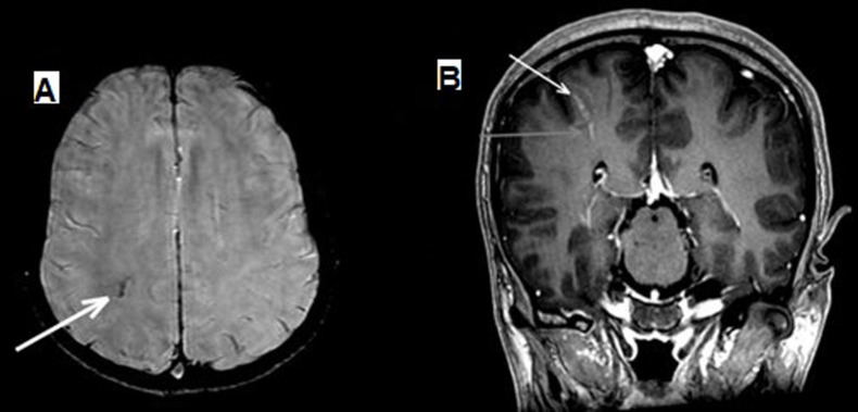
Methods: A record-based study was performed retrospectively in Radiology department at our Hospital in Eastern region of Saudi Arabia, from Jan. 2019-2024. Adult patients for whom MRI brains were conducted for epilepsy work-up with added SWAN (susceptibility weighted angiography) sequence were considered. Imaging proven cases of space occupying lesions in the brain, post-injury, and post-interventional cases were not taken. A venous angioma (or malformation) was documented when a tuft of veins drained to a larger vein (traversing through cortex or reaching under ependymal layer), appeared low signal structure on SWAN image, and enhanced on contrast-enhanced sequence. Consensus reporting was made by 2 experienced neuroradiologists. The usefulness of the SWAN sequence in the detection of venous malformation determined if the visualized abnormality was found to be related to a focus resulting in abnormal waves on the brain electroencephalography. This observation was compared to the accidently found such malformations that were seen in epileptic patients with normal EEGs (control group). Fisher's Exact test was applied and a P-value of <0.05 was taken as statistically significant for an association.
Results: Total number of patients was 114; 65 females (57%) and 49 males (43%), with an average age of 31.4 (range, 15-50). The SWAN found venous angiomas in 34 (29.8%), and 8 were responsible for abnormal electroencephalograms while neither of the 3 accidently detected venous malformations in the control with normal EEGs (p-value=0.001).
Conclusion: An added SWAN sequence with conventional MRI brain imaging for patients with seizures can assist to visualize symptomatic venous angiomas leading to focal seizures.
期刊介绍:
Neurosciences is an open access, peer-reviewed, quarterly publication. Authors are invited to submit for publication articles reporting original work related to the nervous system, e.g., neurology, neurophysiology, neuroradiology, neurosurgery, neurorehabilitation, neurooncology, neuropsychiatry, and neurogenetics, etc. Basic research withclear clinical implications will also be considered. Review articles of current interest and high standard are welcomed for consideration. Prospective workshould not be backdated. There are also sections for Case Reports, Brief Communication, Correspondence, and medical news items. To promote continuous education, training, and learning, we include Clinical Images and MCQ’s. Highlights of international and regional meetings of interest, and specialized supplements will also be considered. All submissions must conform to the Uniform Requirements.
 求助内容:
求助内容: 应助结果提醒方式:


