Athinodoros Athinodorou, Nicolas Israeliantz, Jenna Richardson, Dario Costanza, Jorge Del Pozo, Tobias Schwarz
{"title":"家兔肺气肿的计算机断层扫描及临床表现。","authors":"Athinodoros Athinodorou, Nicolas Israeliantz, Jenna Richardson, Dario Costanza, Jorge Del Pozo, Tobias Schwarz","doi":"10.1111/vru.70063","DOIUrl":null,"url":null,"abstract":"<p><p>Pulmonary emphysema (PE) is a poorly understood condition in rabbits. This retrospective case-control study investigated the CT and clinical findings of rabbits with PE. Institutional archive review identified 724 thoracic CT studies of 529 rabbits, including 76 PE-positive studies of 59/529 rabbits. Twenty-five PE-negative cases were selected randomly as a control group. The mean age of affected rabbits was 9 years (range 5-13 years). Cranial lung lobes were more commonly affected (p < .01). The X-ray attenuation in Hounsfield units (HU) of the emphysematous lung areas (median -905 HU) was significantly lower than in nonemphysematous lung lobes of the case (median -667 HU) and control group (median -652 HU). There was significantly lower X-ray attenuation in peripheral and bullous emphysema than in diffuse emphysema. There was no statistical correlation between clinical lower respiratory signs and PE presence. However, the small portion (n = 6, 10.2%) of affected rabbits with severe respiratory signs, such as open-mouth breathing and cyanotic mucous membranes, all had advanced PE and poor outcome. Secondary changes attributable to PE included pathologic rib fractures in 3 (5.1%) and bulla rupture leading to pneumothorax in 2 (3.4%) rabbits. Of the 15 rabbits with repeat examinations, PE was progressive in 12 (80%) and static in 3 (20%). PE is a common condition in rabbits that is readily detectable with CT. The progressive nature of PE should be considered when detected in asymptomatic rabbits. In rabbits with severe lower respiratory signs, PE should be considered as a potential cause.</p>","PeriodicalId":23581,"journal":{"name":"Veterinary Radiology & Ultrasound","volume":"66 4","pages":"e70063"},"PeriodicalIF":1.5000,"publicationDate":"2025-07-01","publicationTypes":"Journal Article","fieldsOfStudy":null,"isOpenAccess":false,"openAccessPdf":"https://www.ncbi.nlm.nih.gov/pmc/articles/PMC12265035/pdf/","citationCount":"0","resultStr":"{\"title\":\"Computed Tomographic and Clinical Findings in Domestic Rabbits (Oryctolagus cuniculus domesticus) with Pulmonary Emphysema.\",\"authors\":\"Athinodoros Athinodorou, Nicolas Israeliantz, Jenna Richardson, Dario Costanza, Jorge Del Pozo, Tobias Schwarz\",\"doi\":\"10.1111/vru.70063\",\"DOIUrl\":null,\"url\":null,\"abstract\":\"<p><p>Pulmonary emphysema (PE) is a poorly understood condition in rabbits. This retrospective case-control study investigated the CT and clinical findings of rabbits with PE. Institutional archive review identified 724 thoracic CT studies of 529 rabbits, including 76 PE-positive studies of 59/529 rabbits. Twenty-five PE-negative cases were selected randomly as a control group. The mean age of affected rabbits was 9 years (range 5-13 years). Cranial lung lobes were more commonly affected (p < .01). The X-ray attenuation in Hounsfield units (HU) of the emphysematous lung areas (median -905 HU) was significantly lower than in nonemphysematous lung lobes of the case (median -667 HU) and control group (median -652 HU). There was significantly lower X-ray attenuation in peripheral and bullous emphysema than in diffuse emphysema. There was no statistical correlation between clinical lower respiratory signs and PE presence. However, the small portion (n = 6, 10.2%) of affected rabbits with severe respiratory signs, such as open-mouth breathing and cyanotic mucous membranes, all had advanced PE and poor outcome. Secondary changes attributable to PE included pathologic rib fractures in 3 (5.1%) and bulla rupture leading to pneumothorax in 2 (3.4%) rabbits. Of the 15 rabbits with repeat examinations, PE was progressive in 12 (80%) and static in 3 (20%). PE is a common condition in rabbits that is readily detectable with CT. The progressive nature of PE should be considered when detected in asymptomatic rabbits. In rabbits with severe lower respiratory signs, PE should be considered as a potential cause.</p>\",\"PeriodicalId\":23581,\"journal\":{\"name\":\"Veterinary Radiology & Ultrasound\",\"volume\":\"66 4\",\"pages\":\"e70063\"},\"PeriodicalIF\":1.5000,\"publicationDate\":\"2025-07-01\",\"publicationTypes\":\"Journal Article\",\"fieldsOfStudy\":null,\"isOpenAccess\":false,\"openAccessPdf\":\"https://www.ncbi.nlm.nih.gov/pmc/articles/PMC12265035/pdf/\",\"citationCount\":\"0\",\"resultStr\":null,\"platform\":\"Semanticscholar\",\"paperid\":null,\"PeriodicalName\":\"Veterinary Radiology & Ultrasound\",\"FirstCategoryId\":\"97\",\"ListUrlMain\":\"https://doi.org/10.1111/vru.70063\",\"RegionNum\":2,\"RegionCategory\":\"农林科学\",\"ArticlePicture\":[],\"TitleCN\":null,\"AbstractTextCN\":null,\"PMCID\":null,\"EPubDate\":\"\",\"PubModel\":\"\",\"JCR\":\"Q2\",\"JCRName\":\"VETERINARY SCIENCES\",\"Score\":null,\"Total\":0}","platform":"Semanticscholar","paperid":null,"PeriodicalName":"Veterinary Radiology & Ultrasound","FirstCategoryId":"97","ListUrlMain":"https://doi.org/10.1111/vru.70063","RegionNum":2,"RegionCategory":"农林科学","ArticlePicture":[],"TitleCN":null,"AbstractTextCN":null,"PMCID":null,"EPubDate":"","PubModel":"","JCR":"Q2","JCRName":"VETERINARY SCIENCES","Score":null,"Total":0}
Computed Tomographic and Clinical Findings in Domestic Rabbits (Oryctolagus cuniculus domesticus) with Pulmonary Emphysema.
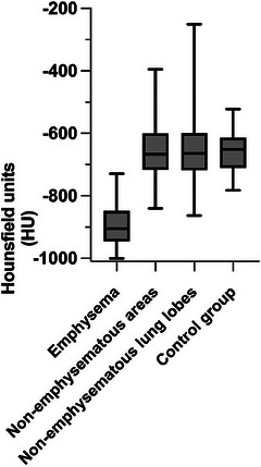
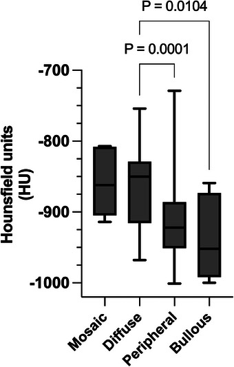
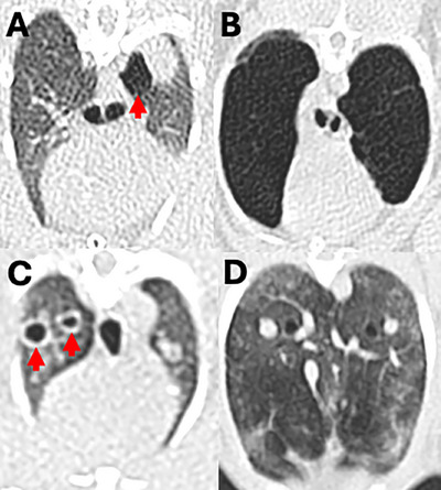
Pulmonary emphysema (PE) is a poorly understood condition in rabbits. This retrospective case-control study investigated the CT and clinical findings of rabbits with PE. Institutional archive review identified 724 thoracic CT studies of 529 rabbits, including 76 PE-positive studies of 59/529 rabbits. Twenty-five PE-negative cases were selected randomly as a control group. The mean age of affected rabbits was 9 years (range 5-13 years). Cranial lung lobes were more commonly affected (p < .01). The X-ray attenuation in Hounsfield units (HU) of the emphysematous lung areas (median -905 HU) was significantly lower than in nonemphysematous lung lobes of the case (median -667 HU) and control group (median -652 HU). There was significantly lower X-ray attenuation in peripheral and bullous emphysema than in diffuse emphysema. There was no statistical correlation between clinical lower respiratory signs and PE presence. However, the small portion (n = 6, 10.2%) of affected rabbits with severe respiratory signs, such as open-mouth breathing and cyanotic mucous membranes, all had advanced PE and poor outcome. Secondary changes attributable to PE included pathologic rib fractures in 3 (5.1%) and bulla rupture leading to pneumothorax in 2 (3.4%) rabbits. Of the 15 rabbits with repeat examinations, PE was progressive in 12 (80%) and static in 3 (20%). PE is a common condition in rabbits that is readily detectable with CT. The progressive nature of PE should be considered when detected in asymptomatic rabbits. In rabbits with severe lower respiratory signs, PE should be considered as a potential cause.
期刊介绍:
Veterinary Radiology & Ultrasound is a bimonthly, international, peer-reviewed, research journal devoted to the fields of veterinary diagnostic imaging and radiation oncology. Established in 1958, it is owned by the American College of Veterinary Radiology and is also the official journal for six affiliate veterinary organizations. Veterinary Radiology & Ultrasound is represented on the International Committee of Medical Journal Editors, World Association of Medical Editors, and Committee on Publication Ethics.
The mission of Veterinary Radiology & Ultrasound is to serve as a leading resource for high quality articles that advance scientific knowledge and standards of clinical practice in the areas of veterinary diagnostic radiology, computed tomography, magnetic resonance imaging, ultrasonography, nuclear imaging, radiation oncology, and interventional radiology. Manuscript types include original investigations, imaging diagnosis reports, review articles, editorials and letters to the Editor. Acceptance criteria include originality, significance, quality, reader interest, composition and adherence to author guidelines.

 求助内容:
求助内容: 应助结果提醒方式:


