Zainab Rustam, Sarah Aman, Nakul Singh, Rose Tan, Amir H Kashani, Peter A Campochiaro
{"title":"Alport综合征的阶梯状/蜂窝状黄斑病变1例报告。","authors":"Zainab Rustam, Sarah Aman, Nakul Singh, Rose Tan, Amir H Kashani, Peter A Campochiaro","doi":"10.1159/000546567","DOIUrl":null,"url":null,"abstract":"<p><strong>Introduction: </strong>Alport syndrome is an inherited disease caused by mutations in COL4A5, COLA3, or COL4A4 resulting in kidney failure, hearing loss, and ocular symptoms. We report a patient with Alport syndrome who has a \"stair-case/honeycomb\" maculopathy, a rare but distinctive finding in this disease.</p><p><strong>Case presentation: </strong>A 53-year-old man with Alport syndrome was referred for gradual decrease in vision. His ocular history was remarkable for intraocular lens implantation secondary to lenticonus in each eye. Fundus photography showed rare white dots in the temporal mid-periphery in each eye and fundus autofluorescence was normal. Optical coherence tomography (OCT) B-scans through the fovea showed irregular thinning of the inner retina with peaks and valleys in the macula of each eye. The ellipsoid zone was intact except for mild patchiness centrally. En face retinal structural OCT angiography (OCTA) images showed a mosaic-like honeycomb pattern in the macular region in both eyes, with hyporeflective depressions in areas of focal retinal atrophy. Retinal OCTA scans showed irregular foveal avascular zone (FAZ) areas with capillaries crossing the FAZ in the left eye, corresponding to islands of preserved retinal tissue. There was predominance of capillaries in the deeper retinal layers centrally.</p><p><strong>Conclusion: </strong>While severe irregular thinning of the macula is not a common feature in Alport syndrome, when it is present in patients who have not been previously diagnosed, particularly in patients with renal disease, it should suggest the diagnosis of Alport syndrome. Its occurrence can be the cause of vision loss which is not commonly associated with Alport central maculopathy.</p>","PeriodicalId":9635,"journal":{"name":"Case Reports in Ophthalmology","volume":"16 1","pages":"496-502"},"PeriodicalIF":0.6000,"publicationDate":"2025-06-24","publicationTypes":"Journal Article","fieldsOfStudy":null,"isOpenAccess":false,"openAccessPdf":"https://www.ncbi.nlm.nih.gov/pmc/articles/PMC12263145/pdf/","citationCount":"0","resultStr":"{\"title\":\"Stair-Case/Honeycomb Maculopathy in Alport Syndrome: A Case Report.\",\"authors\":\"Zainab Rustam, Sarah Aman, Nakul Singh, Rose Tan, Amir H Kashani, Peter A Campochiaro\",\"doi\":\"10.1159/000546567\",\"DOIUrl\":null,\"url\":null,\"abstract\":\"<p><strong>Introduction: </strong>Alport syndrome is an inherited disease caused by mutations in COL4A5, COLA3, or COL4A4 resulting in kidney failure, hearing loss, and ocular symptoms. We report a patient with Alport syndrome who has a \\\"stair-case/honeycomb\\\" maculopathy, a rare but distinctive finding in this disease.</p><p><strong>Case presentation: </strong>A 53-year-old man with Alport syndrome was referred for gradual decrease in vision. His ocular history was remarkable for intraocular lens implantation secondary to lenticonus in each eye. Fundus photography showed rare white dots in the temporal mid-periphery in each eye and fundus autofluorescence was normal. Optical coherence tomography (OCT) B-scans through the fovea showed irregular thinning of the inner retina with peaks and valleys in the macula of each eye. The ellipsoid zone was intact except for mild patchiness centrally. En face retinal structural OCT angiography (OCTA) images showed a mosaic-like honeycomb pattern in the macular region in both eyes, with hyporeflective depressions in areas of focal retinal atrophy. Retinal OCTA scans showed irregular foveal avascular zone (FAZ) areas with capillaries crossing the FAZ in the left eye, corresponding to islands of preserved retinal tissue. There was predominance of capillaries in the deeper retinal layers centrally.</p><p><strong>Conclusion: </strong>While severe irregular thinning of the macula is not a common feature in Alport syndrome, when it is present in patients who have not been previously diagnosed, particularly in patients with renal disease, it should suggest the diagnosis of Alport syndrome. Its occurrence can be the cause of vision loss which is not commonly associated with Alport central maculopathy.</p>\",\"PeriodicalId\":9635,\"journal\":{\"name\":\"Case Reports in Ophthalmology\",\"volume\":\"16 1\",\"pages\":\"496-502\"},\"PeriodicalIF\":0.6000,\"publicationDate\":\"2025-06-24\",\"publicationTypes\":\"Journal Article\",\"fieldsOfStudy\":null,\"isOpenAccess\":false,\"openAccessPdf\":\"https://www.ncbi.nlm.nih.gov/pmc/articles/PMC12263145/pdf/\",\"citationCount\":\"0\",\"resultStr\":null,\"platform\":\"Semanticscholar\",\"paperid\":null,\"PeriodicalName\":\"Case Reports in Ophthalmology\",\"FirstCategoryId\":\"1085\",\"ListUrlMain\":\"https://doi.org/10.1159/000546567\",\"RegionNum\":0,\"RegionCategory\":null,\"ArticlePicture\":[],\"TitleCN\":null,\"AbstractTextCN\":null,\"PMCID\":null,\"EPubDate\":\"2025/1/1 0:00:00\",\"PubModel\":\"eCollection\",\"JCR\":\"Q4\",\"JCRName\":\"OPHTHALMOLOGY\",\"Score\":null,\"Total\":0}","platform":"Semanticscholar","paperid":null,"PeriodicalName":"Case Reports in Ophthalmology","FirstCategoryId":"1085","ListUrlMain":"https://doi.org/10.1159/000546567","RegionNum":0,"RegionCategory":null,"ArticlePicture":[],"TitleCN":null,"AbstractTextCN":null,"PMCID":null,"EPubDate":"2025/1/1 0:00:00","PubModel":"eCollection","JCR":"Q4","JCRName":"OPHTHALMOLOGY","Score":null,"Total":0}
Stair-Case/Honeycomb Maculopathy in Alport Syndrome: A Case Report.
Introduction: Alport syndrome is an inherited disease caused by mutations in COL4A5, COLA3, or COL4A4 resulting in kidney failure, hearing loss, and ocular symptoms. We report a patient with Alport syndrome who has a "stair-case/honeycomb" maculopathy, a rare but distinctive finding in this disease.
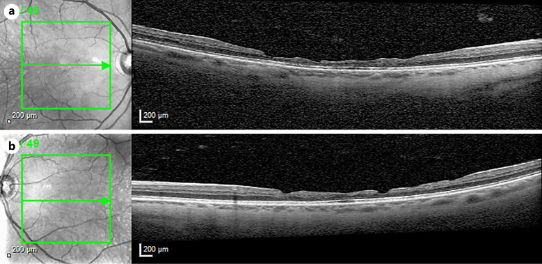
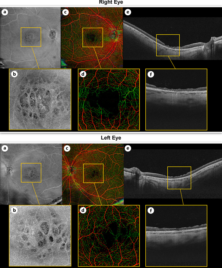
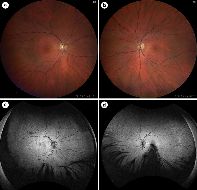
Case presentation: A 53-year-old man with Alport syndrome was referred for gradual decrease in vision. His ocular history was remarkable for intraocular lens implantation secondary to lenticonus in each eye. Fundus photography showed rare white dots in the temporal mid-periphery in each eye and fundus autofluorescence was normal. Optical coherence tomography (OCT) B-scans through the fovea showed irregular thinning of the inner retina with peaks and valleys in the macula of each eye. The ellipsoid zone was intact except for mild patchiness centrally. En face retinal structural OCT angiography (OCTA) images showed a mosaic-like honeycomb pattern in the macular region in both eyes, with hyporeflective depressions in areas of focal retinal atrophy. Retinal OCTA scans showed irregular foveal avascular zone (FAZ) areas with capillaries crossing the FAZ in the left eye, corresponding to islands of preserved retinal tissue. There was predominance of capillaries in the deeper retinal layers centrally.
Conclusion: While severe irregular thinning of the macula is not a common feature in Alport syndrome, when it is present in patients who have not been previously diagnosed, particularly in patients with renal disease, it should suggest the diagnosis of Alport syndrome. Its occurrence can be the cause of vision loss which is not commonly associated with Alport central maculopathy.
期刊介绍:
This peer-reviewed online-only journal publishes original case reports covering the entire spectrum of ophthalmology, including prevention, diagnosis, treatment, toxicities of therapy, supportive care, quality-of-life, and survivorship issues. The submission of negative results is strongly encouraged. The journal will also accept case reports dealing with the use of novel technologies, both in the arena of diagnosis and treatment. Supplementary material is welcomed. The intent of the journal is to provide clinicians and researchers with a tool to disseminate their personal experiences to a wider public as well as to review interesting cases encountered by colleagues all over the world. Universally used terms can be searched across the entire growing collection of case reports, further facilitating the retrieval of specific information. Following the open access principle, the entire contents can be retrieved at no charge, guaranteeing easy access to this valuable source of anecdotal information at all times.

 求助内容:
求助内容: 应助结果提醒方式:


